
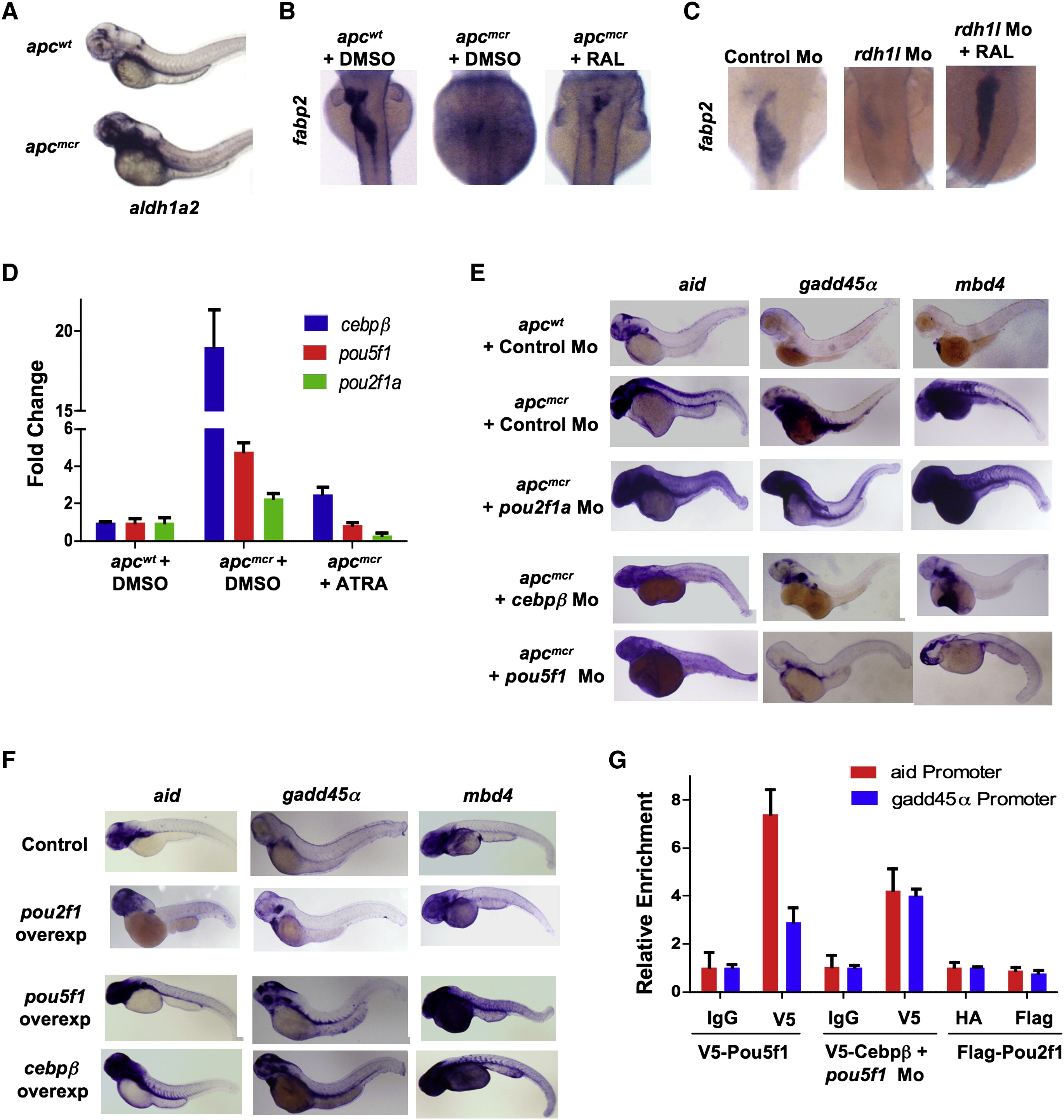
Fig. 3
|
Fig. 3
Pou5f1 and Cebpβ Directly Regulate aid, mbd4, and gadd45α Expression in apcmcr Embryos
(A) Whole-mount in situ staining for aldh1a2 in apcmcr and apcwt at 72 hpf.
(B and C) Whole-mount in situ staining for fabp2 in apcmcr and apcwt embryos (B) or in control morphant and rdh1l morphant embryos (C) (at 72 hpf) treated with vehicle or RAL (2 μM).
(D) RT-PCR for Pou5f1 (Oct4) and Cebpβ in apcmcr and apcwt treated with DMSO or ATRA (1 μM). The y axis shows fold induction normalized to 28S and wild-type DMSO-treated sample.
(E and F) Whole-mount in situ staining for aid, gadd45α, and mbd4 in apcmcr and apcwt injected with control, Pou2f1a, pou5f1, or cebpβ Mo (E) or in wild-type embryos injected with control, Pou2f1, pou5f1, or cebpβ expression plasmid (F).
(G) Graph showing fold enrichment near the aid or gadd45α TSS (containing overlapping Oct and Cebp binding sites) for Cebpβ and Pou5f1 in embryos injected with V5-Cebpβ- (along with Pou5f1 Mo, 80 pg), V5-Pou5f1-, or FLAG-Pou2f1-expressing plasmids. ChIP was performed with antibodies against the tags. Normalization control primers are located not, ~3 kb upstream (region without Cebpβ sites) of the TSS of the Gadd45α gene.
Error bars indicate ± SD. See also Figure S3.
Reprinted from Cell, 142(6), Rai, K., Sarkar, S., Broadbent, T.J., Voas, M., Grossmann, K.F., Nadauld, L.D., Dehghanizadeh, S., Hagos, F.T., Li, Y., Toth, R.K., Chidester, S., Bahr, T.M., Johnson, W.E., Sklow, B., Burt, R., Cairns, B.R., and Jones, D.A., DNA demethylase activity maintains intestinal cells in an undifferentiated state following loss of APC, 930-942, Copyright (2010) with permission from Elsevier. Full text @ Cell