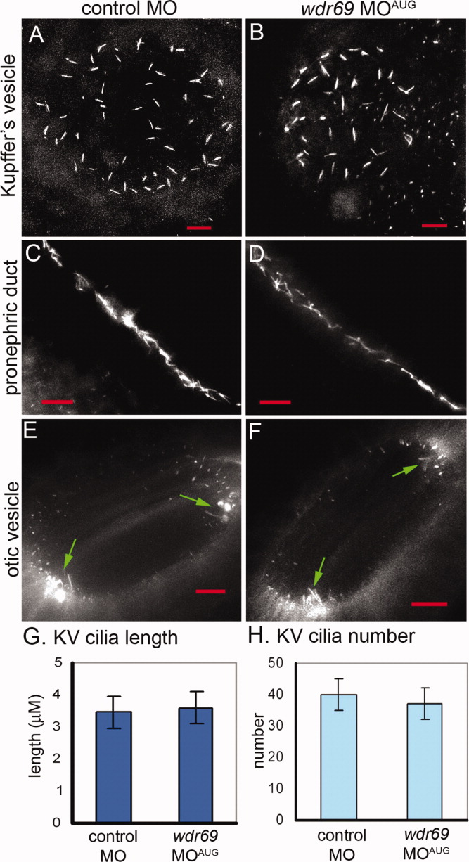
Fig. 4 Cilia length and number is not changed in wdr69 morphants. A-F: Cilia were visualized by immunofluorescent staining with acetylated tubulin antibodies. Cilia in embryos injected with wdr69 MOAUG appeared similar to cilia injected with control MO in Kupffer′s vesicle (KV; A,B), pronephric ducts (C,D) and otic vesicles (arrows in E,F point to motile tether cilia). All red scale bars represent 10 μM. G,H: Measurements of KV cilia at six to eight somite stage (SS) showed no significant differences in the average length (G) or number (H) between wdr69 MOAUG (n = 17) and control (n = 21) morphants. Error bars = one standard deviation. MO, morpholino oligonucleotides.
Image
Figure Caption
Figure Data
Acknowledgments
This image is the copyrighted work of the attributed author or publisher, and
ZFIN has permission only to display this image to its users.
Additional permissions should be obtained from the applicable author or publisher of the image.
Full text @ Dev. Dyn.

