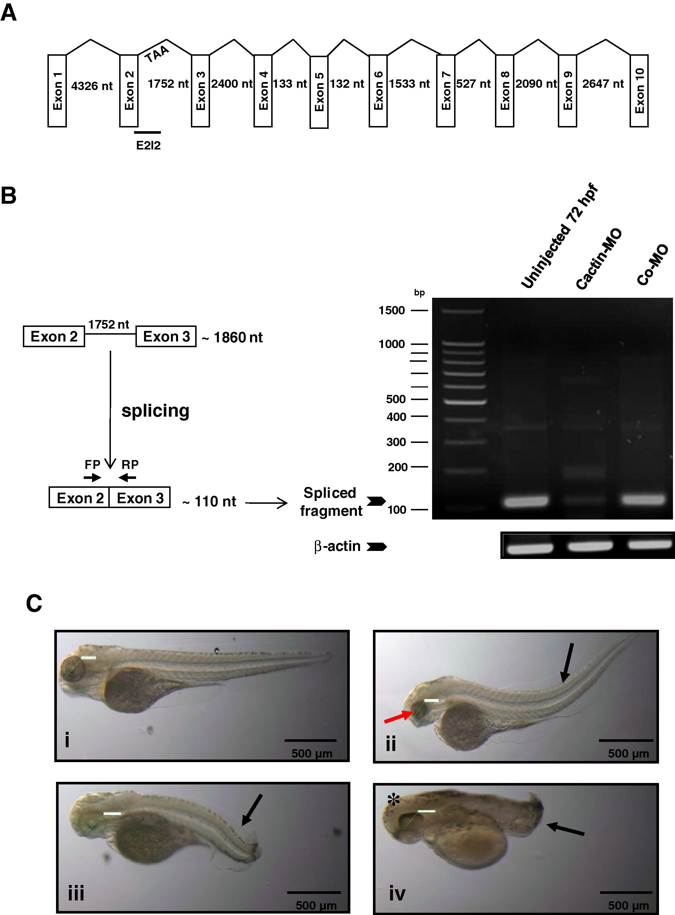
Fig. 3 Knockdown of zCactin by specific morpholino antisense oligonucleotide causes developmental abnormalities. (A) Schematic representation shows the genomic organisation of the zCactin gene (not in scale). Splice site targeted by E2I2 morpholino is shown. Stop codon in intron 2 is indicated (TAA). (B) The efficacy of the morpholino was validated by RT-PCR using a forward primer (FP) and reverse primer (RP) as indicated in uninjected embryos, Cactin morpholino (Cactin-MO) and control morpholino (Co-MO) injected embryos. The PCR products of the spliced form are illustrated together with a PCR fragment of the β-actin housekeeping gene. (C) Representative images of embryos (72 hpf) injected with control morpholino (Co-MO) (i) or Cactin morpholino (Cactin-MO) (ii–iv) (300μM) are demonstrated and are representative of three independent experiments. Each experiment comprised of two groups of embryos injected with each morpholino (98, 112 and 74 embryos for Cactin-MO and 90, 152 and 88 embryos for Co-MO). Black arrows indicate bent body characteristics, the red arrow points to smaller eye size and the asterisk shows the more rounded head phenotype. Eye size was measured (green line); for Co-MO: 736.42μm; for Cactin-MO: 558.77, 597.26 and 808.78 μm. Images were acquired using a Zeiss Axioplan II microscope and processed using Adobe Photoshop.
Reprinted from Gene expression patterns : GEP, 10(4-5), Atzei, P., Yang, F., Collery, R., Kennedy, B.N., and Moynagh, P.N., Characterisation of expression patterns and functional role of Cactin in early zebrafish development, 199-206, Copyright (2010) with permission from Elsevier. Full text @ Gene Expr. Patterns

