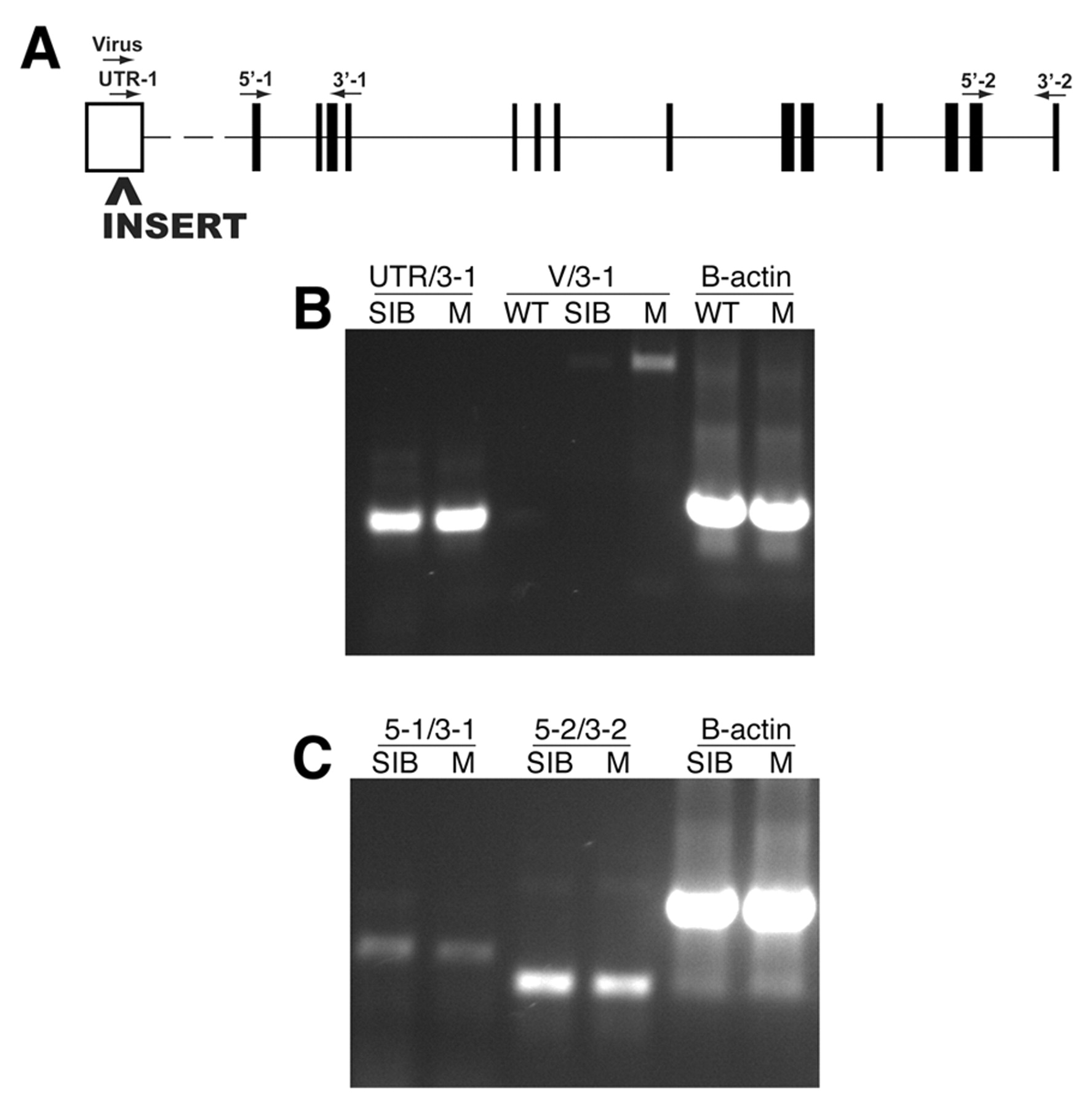
Fig. 4 Molecular analysis of the garthi3526b locus. (A) The approximate position of the proviral insertion and RT-PCR primers mapped onto a schematic of the mutated gene (www.ensembl.org/Danio_rerio/). The proviral insertion is located in the predicted 5′ UTR. Amplification of the region between the UTR and exon 4 (B, UTR/3-1), the region between exons 1 and 4 (C, 5-1/3-1), or the region between exons 13 and 14 (C, 5-2/3-2) all indicate that the overall levels of gart transcripts are unaffected in the homozygous mutant embryos. Amplification from the 3′ end of the proviral insert to exon 4 (B, V/3-1) in homozygous gart mutants indicates that a portion of the proviral insert is transcribed and retained in the resulting mRNA. WT, wild-type AB; SIB, wild-type sibling mixture (+/+ and +/-); M, homozygous mutant.
Image
Figure Caption
Acknowledgments
This image is the copyrighted work of the attributed author or publisher, and
ZFIN has permission only to display this image to its users.
Additional permissions should be obtained from the applicable author or publisher of the image.
Full text @ Development

