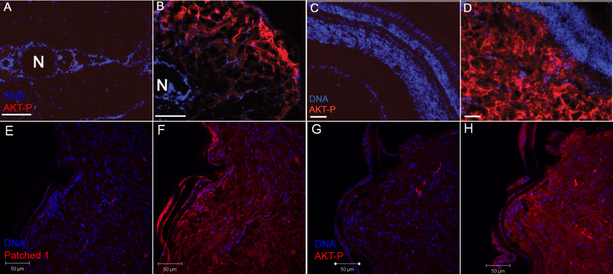
Fig. 3 Elevated phospho-AKT1 and Patched 1 levels in zebrafish tumors. A, B, a 3-week-old transgenic fish with trunk tumor (B) and its age-matched tumor-free fish (A). C, D, a 12-week-old fish with an eye tumor (D) and its age-matched tumor free fish (C). Immunofluorescence was done on cryosections for the above fish. E-H, an astrocytoma from a double transgenic fish showed elevated levels of both Patched 1 (F) and phosphorylated AKT (H). Immunofluoresence was done on paraffin sections for this tumor. Negative controls for Patched 1 (E) and phosphorylated AKT (G) were treated the same way except no primary antibodies were added. N, Notochord; AKT-P, phosphorylated AKT. Scale bars, 100 μm for A-D, 50 μm for E-H.
Image
Figure Caption
Figure Data
Acknowledgments
This image is the copyrighted work of the attributed author or publisher, and
ZFIN has permission only to display this image to its users.
Additional permissions should be obtained from the applicable author or publisher of the image.
Open Access.
Full text @ Mol. Cancer

