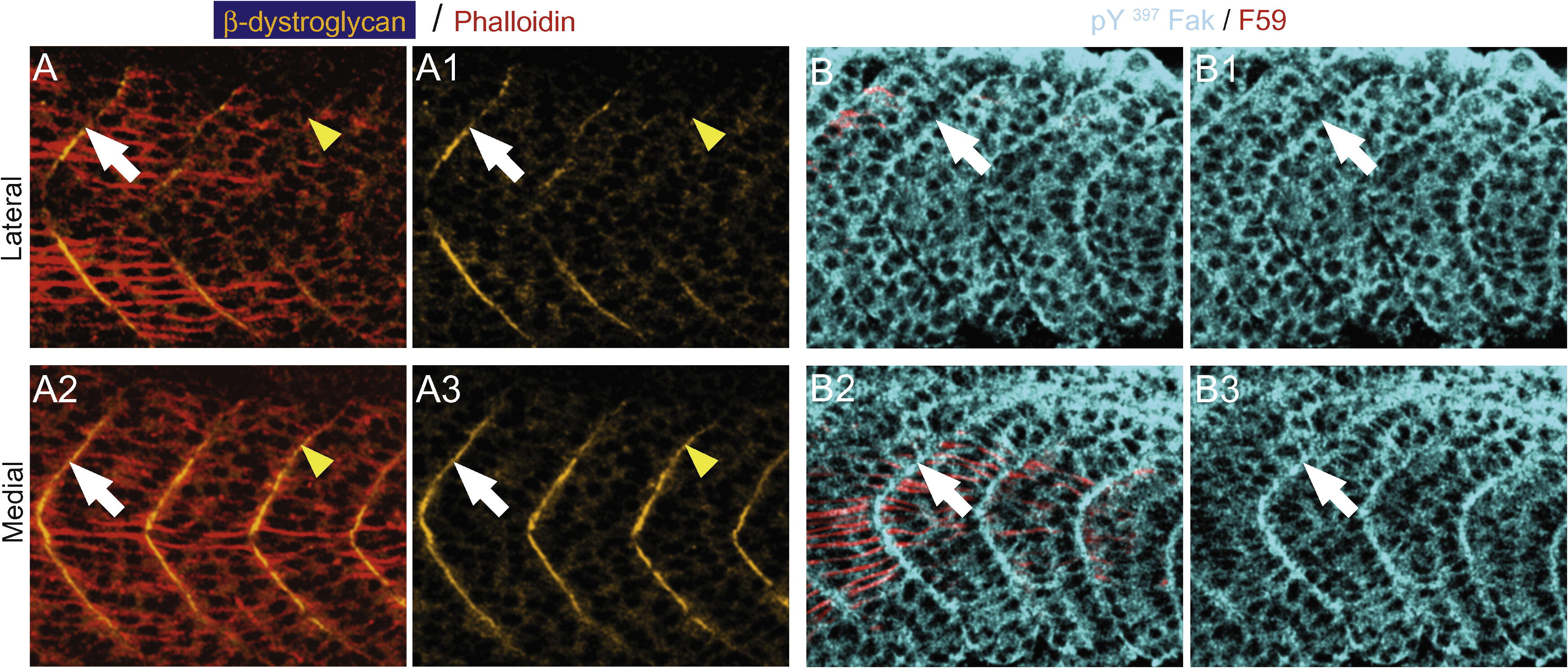
Fig. 3 β-Dystroglycan and pY397 FAK concentrate at the myoseptal tendon medial to migrating slow muscle fibers. All panels are ApoTome images, side views, anterior left, dorsal top, of the transition region, where slow muscle is migrating, in 19–21 somite embryos. Phalloidin, denoting actin, is red in panel A and F59, denotes slow-twitch muscle in red for panel B. Panels numbered 1 and 3 show only β-dystroglycan and pY397 FAK, respectively. (A) Lateral to migrating slow fibers, β-dystroglycan (green) does not concentrate at the boundary (A, A1, yellow arrowheads) but does concentrate slightly anteriorly where slow-twitch muscle has migrated (white arrows, A, A1). β-Dystroglycan concentrates at the boundary medial to migrating slow fibers (A2, A3, note robust concentration adjacent to yellow arrowheads where β-dystroglycan was not concentrated laterally). (B) Similarly, pY397 FAK concentration at the boundary is increased medial and adjacent to slow muscle fibers (B2, B3, white arrowheads).
Reprinted from Gene expression patterns : GEP, 9(1), Snow, C.J., and Henry, C.A., Dynamic formation of microenvironments at the myotendinous junction correlates with muscle fiber morphogenesis in zebrafish, 37-42, Copyright (2009) with permission from Elsevier. Full text @ Gene Expr. Patterns

