
|
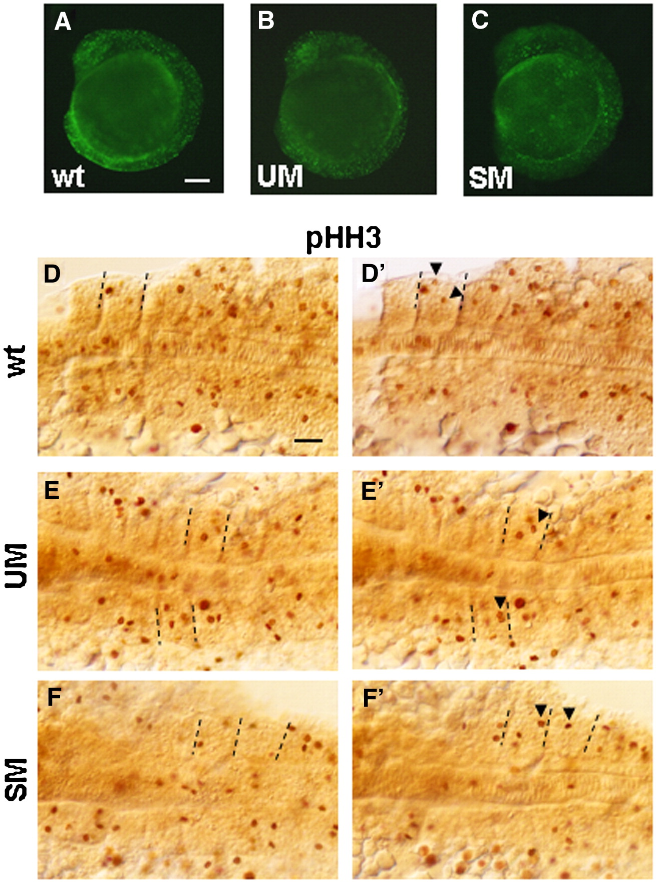
Fig. S2 Staining with acridine orange and anti-phosphohistone H3 Ab at 9 s. (A–C) Fluorescent images of control and morphant embryos stained with acridine orange. Dorsal (D, E, F) and ventral (D′, E′, F′) focal planes of flat mounted embryos after immunohistochemistry with anti-phosphohistone H3 Ab. Posterior 6–7 somites are shown. Equal numbers (n = 10) of control embryos and morphants were stained. Arrowheads in D′, E′, F′ indicate phosphohistone H3-positive cells not clearly seen in D, E, F. Dashed lines mark the borders of somites containing these cells. Stained cells were counted in all 9 somites of 3 embryos for each condition (control, UM and SM morphants) and results were summarized in Table 1. No change in phoshohistone-positive cells in notochord was found. Scale bars, 100 μm.
Reprinted from Developmental Biology, 323(2), Bessarab, D.A., Chong, S.W., Srinivas, B.P., and Korzh, V., Six1a is required for the onset of fast muscle differentiation in zebrafish, 216-228, Copyright (2008) with permission from Elsevier. Full text @ Dev. Biol.