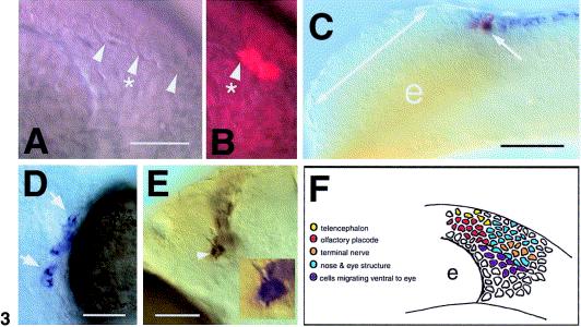
Fig. 3 Localization of GnRH progenitors in the cranial neural crest (CNC). (A) Nomarski image of the border (arrowheads) apparent within premigratory crest at 6-7 somites, anterior to the left. (B) Two cells labeled with lineage tracers within the premigratory CNC (arrowhead with asterisk), dorsal to the border. (C) Lateral view of embryo, anterior to the left. Two cells were labeled (arrow, red) within the fkh6 domain (blue). The double-headed arrow indicates the region of the developing olfactory field; e, eye. (D) Ventral view of whole-mount head with clones of cells (arrows) in the eye sclera. (E) Ventral view of whole-mount head showing cells in the TN (arrowhead) positive for GnRH. Inset is of cell indicated by arrowhead. (F) Summary diagram of lineage tracing. Diagram is a lateral view (see A) of the embryo with cells color-coded as to clone types. Each colored cell indicates one of two to three possible cells labeled in each preparation, thus representing the location of the label in the developing embryo. Scale bars: (A, B) 35 μm; (C) 75 μm; (D) 40 μm; (E) 35 μm; inset cell body, 12 μm.
Reprinted from Developmental Biology, 257(1), Whitlock, K.E., Wolf, C.D., and Boyce, M.L., Gonadotropin-releasing hormone (gnrh) cells arise from cranial neural crest and adenohypophyseal regions of the neural plate in the zebrafish, Danio rerio, 140-152, Copyright (2003) with permission from Elsevier. Full text @ Dev. Biol.

