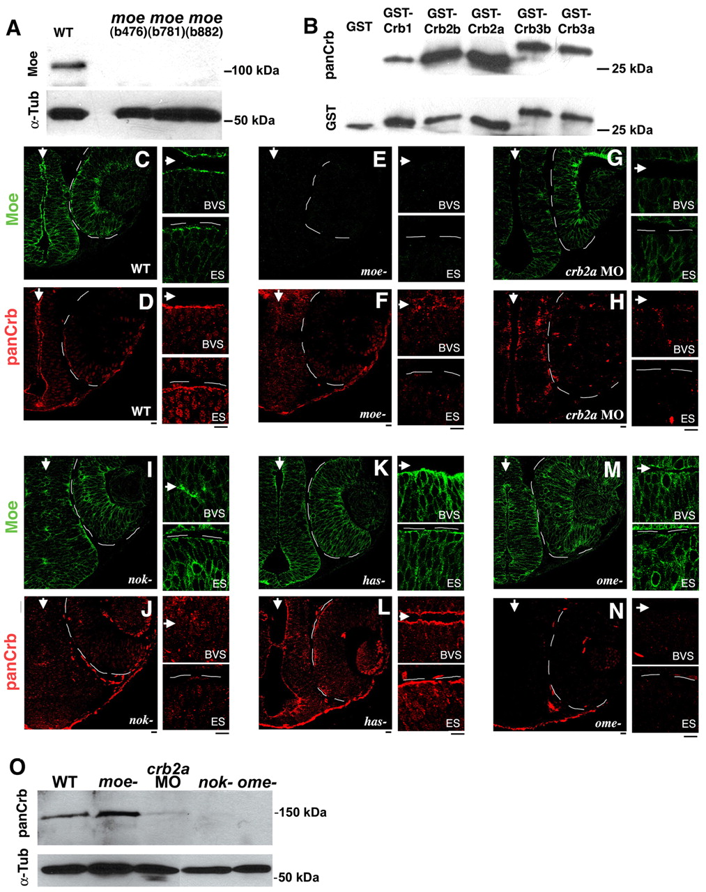
Fig. 2 Localization of Moe and Crumbs proteins in embryonic brain and eye. (A) Western blot of wild type, moeb476, moeb781, moeb882 3d larvae probed with rabbit anti-Moe. Anti-Moe recognizes a protein of approximately 110 kDa in wild-type extracts that is absent in moe mutants. The blot was reprobed with anti-α-Tubulin as a loading control. (B) Anti-CRB3 recognizes the intracellular domains of all zebrafish Crumbs proteins. Western blot of GST, GST-Crb1intra, GST-Crb2aintra, GST-Crb2bintra, GST-Crb3aintra, GST-Crb3bintra probed with anti-CRB3 antibodies. The blot was reprobed with anti-GST as a loading control. (C-N) Moe (green) and panCrb (red) labeling in 30 hpf brain and eye in wild-type (C,D), moeb781 (E,F), crb2a morphant (G,H), nok (I,J), has (K,L) and ome (M,N) embryos. Arrows indicate brain ventricles and eyes are outlined. High magnification of the apical ventricular surface of the brain (BVS) and eye/retinal surface (ES) are shown to the right. Scale bars: 10 μm. (O) Western blot of 30 hpf wild-type, moeb781, crb2a morphant, nok and ome embryos probed with anti-panCrb antibodies. One-fifth of the material loaded for the panCrb Western was probed with anti-α-Tubulin to show relative loading amounts.
Image
Figure Caption
Figure Data
Acknowledgments
This image is the copyrighted work of the attributed author or publisher, and
ZFIN has permission only to display this image to its users.
Additional permissions should be obtained from the applicable author or publisher of the image.
Full text @ Development

