
|
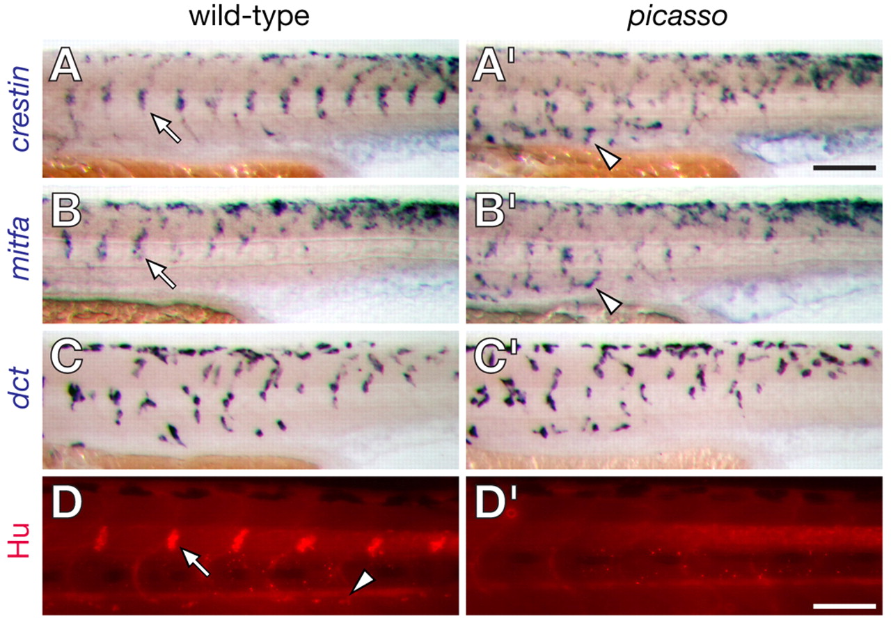
Fig. 11 picasso mutant embryos have defects in neural crest morphogenesis. (A,A′) crestin+ cells form segmentally arranged clusters prior to ganglion formation in wild-type embryos (e.g. arrow in A) but appear to migrate past their normal target sites in picasso mutant embryos (e.g. arrowhead in A′). (B,B′) mitfa+ cells in wild-type exhibit some segmental patterning and a defect in picasso mutants similar to that of crestin+ cells. (C,C′) dct+ melanoblast distributions do not differ consistently between wild-type and picasso mutant embryos. (D,D′) Anti-Hu immunoreactivity shows dorsal root ganglia (e.g. arrow in D) and sympathetic ganglia (e.g. arrowhead in D) in wild-type larvae but their absence in picasso mutants. Scale bar: in A′, 800 μm for A-C′; in D′, 600 μm for D-D′.