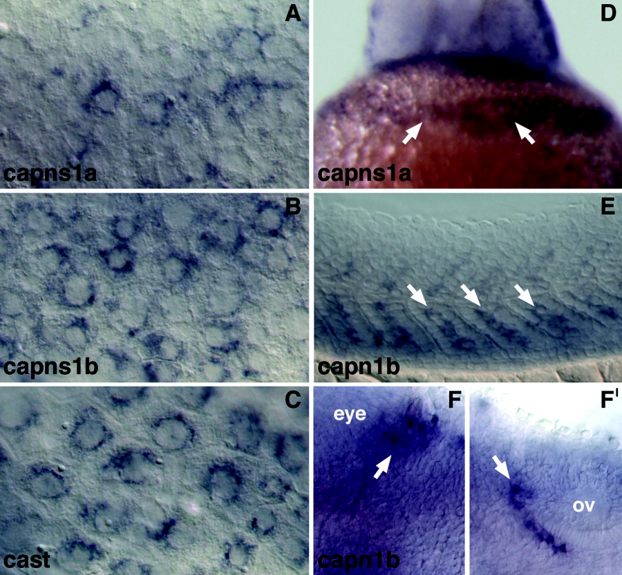
Fig. 6 Tissue-specific expression of capns1a, capns1b, capn1b, and cast by whole-mount in situ hybridization. A-C: Higher magnification views of the enveloping layer (EVL) expression of capns1a (A), capns1b (B), and cast (C) in flat-mounted 4-6 somite stage embryos shown in Figure 5R,V,Z. D: Ventral view of 1 day postfertilization (dpf) embryo shown in Figure 5S, showing expression of capns1a in the hatching gland (arrows). E-F′: Higher magnification views of capn1b expression in medial somites at 1 dpf (E, arrows) and two groups of bilateral (left side shown) hindbrain neuron clusters at 2 dpf (F,F′, arrows). Embryo in E is shown in lateral view and embryos in F and F′ are viewed dorsally. All embryos E-F′ are oriented with anterior to the left. Position of the eye and otic vesicle (ov) are indicated in F and F′, respectively.
Image
Figure Caption
Figure Data
Acknowledgments
This image is the copyrighted work of the attributed author or publisher, and
ZFIN has permission only to display this image to its users.
Additional permissions should be obtained from the applicable author or publisher of the image.
Full text @ Dev. Dyn.

