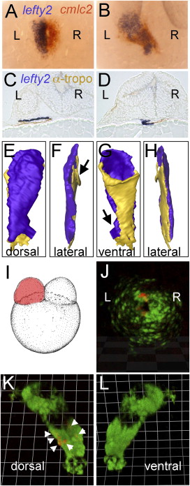
Fig. 2 lefty2 Expression and Lineage Tracing Reveals a Dorsal-Ventral Orientation of the Left-Right Regions of the Heart Field, Respectively, in the Linear Heart Tube (A and B) Double ISH with cmlc2 (red) and lefty2 (blue) antisense riboprobes at 23-somite stage (A) and 24 hpf (B). Dorsal views with anterior to the top. (C and D) Serial section and 3D reconstruction (E–H) of an ISH embryo with lefty2 probe counterstained by using an anti-tropomyosin antibody at the level of the venous pole of the heart tube (C) and the arterial pole (D). (E–H) 3D reconstruction with blue representing lefty2-positive tissue and yellow representing tropomyosin-positive tissue. (E) Dorsal view with venous pole to the top and arterial pole to the bottom. Arrows in (F) and (G) indicate regions at the poles where some torsion of the tube is visible. (I–L) Lineage tracing of CPCs from the left or right cardiac field. (I) CPCs were mosaically labeled in the left or right cardiac field by injection of cmlc2:mRFP DNA into one cell at the two-cell stage. (J) In the cardiac cone, mRFP-expressing CPCs were observed on the left side, and these cells were later found to occupy the dorsal part (K, arrowheads) of the linear heart tube with no mRFP cells observable on the ventral side of the tube (L).
Reprinted from Developmental Cell, 14(2), Smith, K.A., Chocron, S., von der Hardt, S., de Pater, E., Soufan, A., Bussmann, J., Schulte-Merker, S., Hammerschmidt, M., and Bakkers, J., Rotation and asymmetric development of the zebrafish heart requires directed migration of cardiac progenitor cells, 287-297, Copyright (2008) with permission from Elsevier. Full text @ Dev. Cell

