Fig. 6
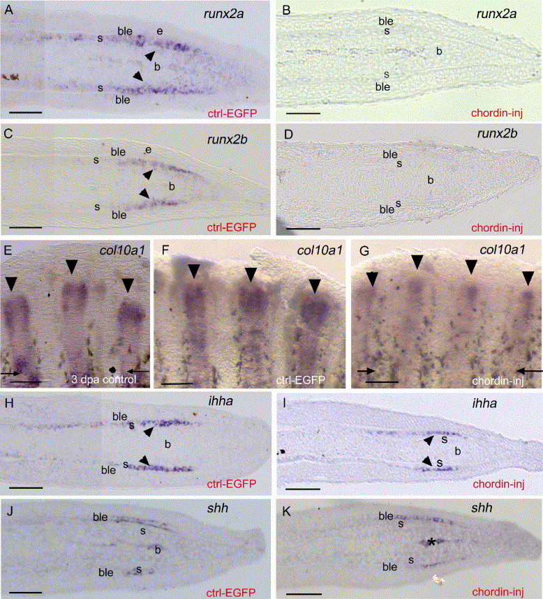
Fig. 6 Gene expression analysis using in situ hybridization on whole-mount and longitudinal sections of EGFP (A, C, F, H, J) and chordin-injected rays (B, D, G, I, K) at 4 dpa/1 dpi. (A-D) runx2a and runx2b are both expressed within newly differentiating scleroblasts (arrowheads in panels A and C) in control regenerates and are both downregulated following chordin injection (B, D). (E-F) col10a1 expression on whole-mount 3 dpa regenerate (E) and 4 dpa regenerate 1 day following EGFP construct injection (F). At 3 dpa col10a1 is already strongly expressed in the entire fin ray and presents a stronger expression in the distal part of the regenerate. At 4 dpa/1 dpi, in the EGFP-injected fin rays, a similar strong expression is observed with a broad domain in the distal part of the regenerate. (G) In contrast, in the chordin-injected rays, col10a1 expression at 4 dpa/1 dpi is reduced and observed in a more restricted domain in the distal part of the regenerate. (H, I) ihha expression in the distal scleroblasts (arrowheads) of EGFP-injected rays (H) is unaffected in chordin-injected rays (I). (J, K) shh expression in the basal epidermal layer is unchanged in both EGFP (J) and chordin-injected rays (K). Asterisks in panels J and K indicate blood vessels. b, blastema; ble, basal layer of the epidermis; s, scleroblasts. Distal part of the regenerate is to the right (A-D, H-K) and to the top (E-G). Arrows indicate level of amputation. Arrowheads show expression of col10a1 in each ray in panels E-G. Scale bars: (A-D, H-K) = 25 μm, (E-G) = 100 μm.
Reprinted from Developmental Biology, 299(2), Smith, A., Avaron, F., Guay, D., Padhi, B.K., and Akimenko, M.A., Inhibition of BMP signaling during zebrafish fin regeneration disrupts fin growth and scleroblast differentiation and function, 438-454, Copyright (2006) with permission from Elsevier. Full text @ Dev. Biol.

