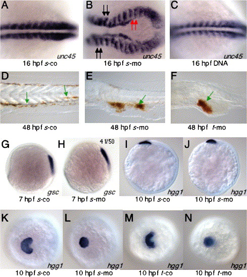
Fig. 5 Sid4 function during development of ventral and prechordal mesoderm. (A) In s-co embryos, somitic mesoderm develops normally. (B) Somite shape and convergence of caudal somitic mesoderm are disrupted in s-mo embryos. Somites anterior (black arrows) to and involved in (red arrows) the convergence defect are similarly malformed. (C) Somite development was normal in embryos injected with 25 pg/nl of sid4 expression DNA. (D–F) Red blood cells stained with o-dianisidine. (D) Red blood cells can be seen within the vasculature of 24 hpf control embryos (green arrows). (E) In s-mo and (F) t-mo embryos, isolated red blood cells are seen in the proximity of blood vessels (green arrows), but the vast majority remain clumped within the abnormally expanded inner cell mass. (G) Anterior migration of gsc-expressing cells was similar in 7 hpf s-co and (H) s-mo embryos. (I) The anterior position of hgg1 hybridization beneath the eye primordium of 10 hpf s-co and (J) s-mo embryos was also normal. (K) Lateral migration of hatching gland cells is normal in s-co and (M) t-co morphants. (L) In both 10 hpf s-mo and (N) t-mo morphants, hatching gland cells do not spread but remain in the midline. Embryonic stages and treatment are indicated below each panel. In situ probes are indicated at the lower right of each panel. Anterior is to the left. A–J are lateral views. K–N are anterodorsal views.
Reprinted from Developmental Biology, 282(1), diIorio, P.J., Runko, A., Farrell, C.A., and Roy, N., Sid4: A secreted vertebrate immunoglobulin protein with roles in zebrafish embryogenesis, 55-69, Copyright (2005) with permission from Elsevier. Full text @ Dev. Biol.

