
Fig. 5
|
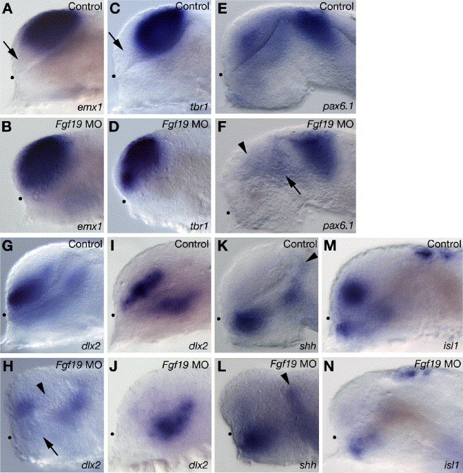
Fig. 5 Telencephalic and diencephalic gene expression in the Fgf19 MO-injected embryos. Embryos were injected with control Fgf19 MO (A, C, E, G, I, K and M) and Fgf19 MO (B, D, F, H, J, L and N). (A–D) The pallial telencephalic markers, emx1 (A and B) and tbr1 (C and D) were expressed in both the pallial and subpallial telencephalon in the Fgf19 MO-injected embryos at 25 hpf. Arrows in panels A and C indicate the subpallial telencephalon that is negative for emx1 or tbr1. (E and F) In the Fgf19 MO-injected embryos, pax6.1 expression in the posterior ventral thalamus (arrow) and dorsal thalamus was unaltered, while the expression in the telencephalon (arrowhead) was reduced at 25 hpf. (G and H) In the Fgf19 MO-injected embryos, dlx2 expression was completely lost in both the posterior ventral telencephalon (arrowhead) and anterior ventral thalamus (arrow) at 25 hpf. (I and J) In the Fgf19 MO-injected embryos, dlx2 expression in the ventral thalamus was unaltered, while the expression in the telencephalon was lost at 16 hpf. (K and L) shh was expressed in similar domains in the control Fgf19 MO-injected and Fgf19 MO-injected embryos at 25 hpf. Arrowheads indicate the zli. (M and N) In the Fgf19 MO-injected embryos, isl1 expression is virtually undetectable in the subpallial telencephalon and reduced in the ventral thalamus and epiphysis at 25 hpf. Lateral views with anterior to the left and dorsal to the top. Dots indicate the boundary between the telencephalon and ventral diencephalon.
Reprinted from Developmental Biology, 288(1), Miyake, A., Nakayama, Y., Konishi, M., and Itoh, N., Fgf19 regulated by Hh signaling is required for zebrafish forebrain development, 259-275, Copyright (2005) with permission from Elsevier. Full text @ Dev. Biol.