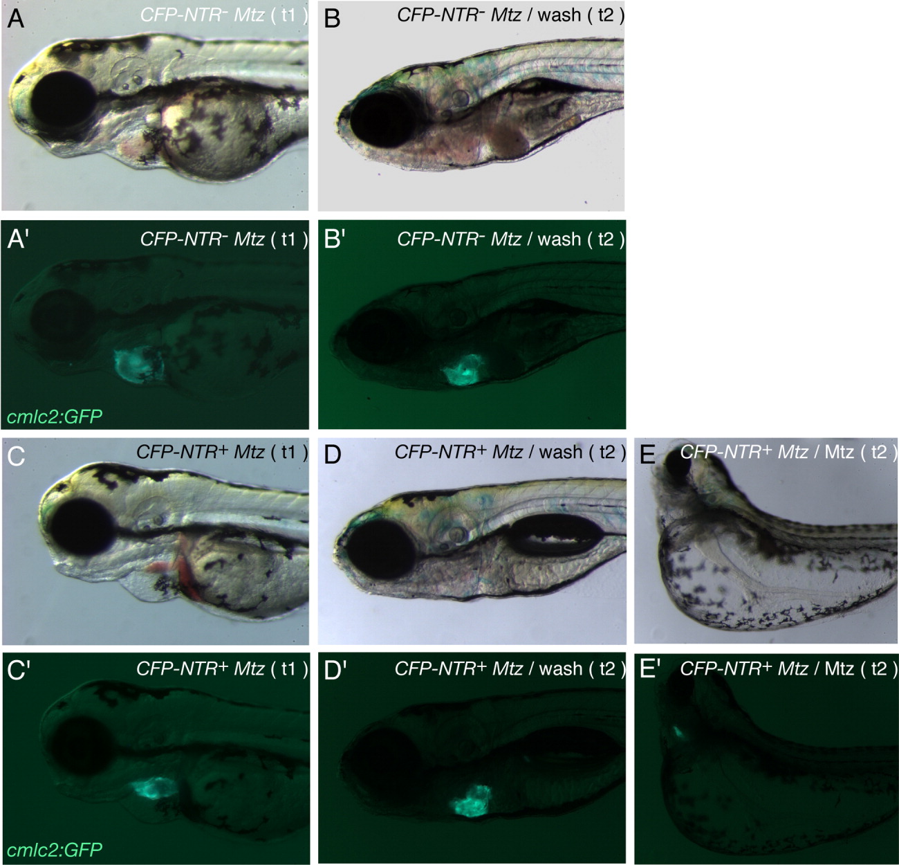
Fig. 3 Functional heart tissue recovery after Nitroreductase/Metronidazole (NTR/Mtz) -mediated cell ablation. A-D: Brightfield images of a control Tg(cmlc2:GFP) larva (A,B) and Tg(cmlc2:CFP-NTR)s890; Tg(cmlc2:GFP) larva (C,D) exposed to 10 mM Mtz at 48 hours postfertilization (hpf), after 24 hr incubation in Mtz (t1; A,C) and after 96 hr washing-out in Mtz-free medium (t2; B,D). E: Brightfield image of Tg(cmlc2:CFP-NTR)s890; Tg(cmlc2:GFP) larva incubated in 10 mM Mtz continuously from 48 to 168 hpf (t2). A′,B′,C′,D′,E′: Fluorescence images showing Tg(cmlc2:GFP) expression (green) in the larvae above (A′ corresponding to A, B′ to B, C′ to C, D′ to D, and E′ to E). Tg(cmlc2:CFP-NTR)s890; Tg(cmlc2:GFP) larvae whose heart tissue was severely damaged as a result of exposure to Mtz for 24 hr (C, C′) were able to replace damaged tissue by functional tissue upon removal of the drug: after 96 hr in Mtz-free medium, the larvae showed a morphologically wild-type heart (D′) and no blood pooling (D). Tg(cmlc2:CFP-NTR)s890; Tg(cmlc2:GFP) larvae continuously exposed to Mtz for 120 hr showed enhanced cardiac and body phenotype defects: curved body, large yolk edema, small eyes, enlarged eye pockets (E), and collapsed, nonfunctional heart chambers of significantly reduced size (E′).
Image
Figure Caption
Acknowledgments
This image is the copyrighted work of the attributed author or publisher, and
ZFIN has permission only to display this image to its users.
Additional permissions should be obtained from the applicable author or publisher of the image.
Full text @ Dev. Dyn.

