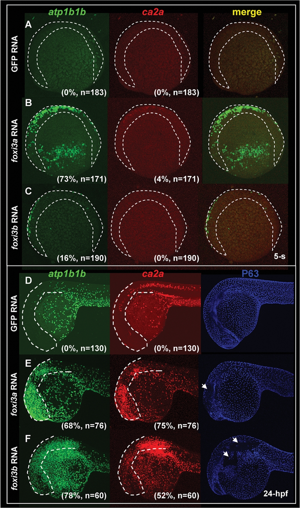
Fig. 7 Misexpression of Either foxi3a or foxi3b in a Wild-type Background is Sufficient to Ectopically Generate Precocious Differentiated Ionocytes in the Epidermis. (A–C) Fluorescent double in situ hybridization with atp1b1b (green) and ca2a (red) probes on wild-type embryos, foxi3a mRNA-injected embryos, and foxi3b mRNA-injected embryos aged at the 5-somite (5-s) stage. For wild-type embryos, normal Na+,K+-ATPase rich cell (NaRC) (atp1b1b+) and H+-ATPase rich cell (HRC) (ca2a+ and atp1b1b+) differentiation was not activated until the 14-s and 18-s stages, respectively. However, when foxi3a mRNA was misexpressed in wild-type embryos, it was sufficient to promote the precocious differentiation of both NaRCs and HRCs in the ventral ectoderm and ectopic sites (labeled by a dotted line) at the 5-s stage. For foxi3b mRNA-misexpression, it is only sufficient to promote the precocious differentiation of NaRCs in the ventral ectoderm and ectopic sites (label by dotted line) at the 5-s stage. (D–F) Triple labeling of atp1b1b (green), ca2a (red), and P63 (blue) on wild-type embryos, foxi3a mRNA-injected embryos, and foxi3b mRNA-injected embryos aged at 24 hours post-fertilization (hpf). For wild-type embryos (D), both NaRCs and HRCs seldom appeared on the cephalic ectoderm (highlighted by a dotted line). However, when either foxi3a mRNA (E) or foxi3b mRNA (F) was misexpressed in wild-type embryos, it was sufficient to generate the ectopic NaRCs and HRCs on the cephalic ectoderm. Ectopic epidermal ionocytes occur at the expanse of epidermal stem cell fate, since some regions with strong atp1b1b and ca2a expression completely lacked P63 expression (indicated by arrows).
Image
Figure Caption
Acknowledgments
This image is the copyrighted work of the attributed author or publisher, and
ZFIN has permission only to display this image to its users.
Additional permissions should be obtained from the applicable author or publisher of the image.
Full text @ PLoS One

