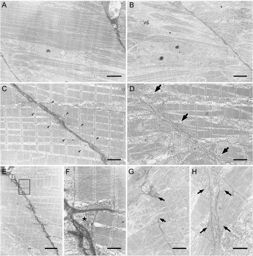
Fig. 8 Electron micrographs of sagittal sections through somites of control MO (A, C, E, and F) and periostin MO (B, D, G, and H)-injected embryos at 48 hpf. The myotomes of control MO-injected embryos are occupied with striated muscle fibers (A and E), and ordered staircase-like structures are seen at the place where a myoseptum and myofibers contact one another (C, arrowheads), whereas the triangular areas are not filled with actin–myosin filaments (arrowheads in C and asterisk in F). Higher magnification of the area boxed (E) shows small protrusions that extend from the myosepta and a triangular area occupied by granular substances (F, asterisk). Conversely, in myotomes of periostin morphants, immature myoblasts are included (B), and striated myofibers of various widths show more frequent branching and merging (D). Moreover, the staircase-like structure is not observed (arrows in D), and the myoseptum is partly disrupted (arrows in G). At the contact points between a myoseptum and myofibers, fibrils are appeared in the whole triangular area and they connect the myoseptum with the z-discs (arrows in H). Scale bars: A and B, 6 μm; C and D, 3 μm; E and G, 4 μm; F and H, 0.7 μm.
Reprinted from Developmental Biology, 267(2), Kudo, H., Amizuka, N., Araki, K., Inohaya, K., and Kudo A., Zebrafish periostin is required for the adhesion of muscle fiber bundles to the myoseptum and for the differentiation of muscle fibers, 473-487, Copyright (2004) with permission from Elsevier. Full text @ Dev. Biol.

