Image
Figure Caption
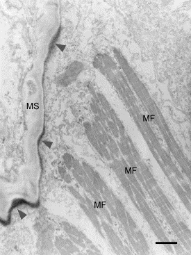
Fig. 4 Immunoelectron microscopic image of periostin in myoseptum of 48-hpf embryos. Myofibrils and myoseptum are obliquely positioned with respect to each other. Periostin immunoreactivity (black color, arrowheads) is observed along the surface of the myoseptum. In addition, the immunoreactivity is concentrated at the intersection of myosepta and extended lines of myofibers. Abbreviations: MF, myofibers; MS, myoseptum. Scale bar: 5 μm.
Acknowledgments
This image is the copyrighted work of the attributed author or publisher, and
ZFIN has permission only to display this image to its users.
Additional permissions should be obtained from the applicable author or publisher of the image.
Reprinted from Developmental Biology, 267(2), Kudo, H., Amizuka, N., Araki, K., Inohaya, K., and Kudo A., Zebrafish periostin is required for the adhesion of muscle fiber bundles to the myoseptum and for the differentiation of muscle fibers, 473-487, Copyright (2004) with permission from Elsevier. Full text @ Dev. Biol.

