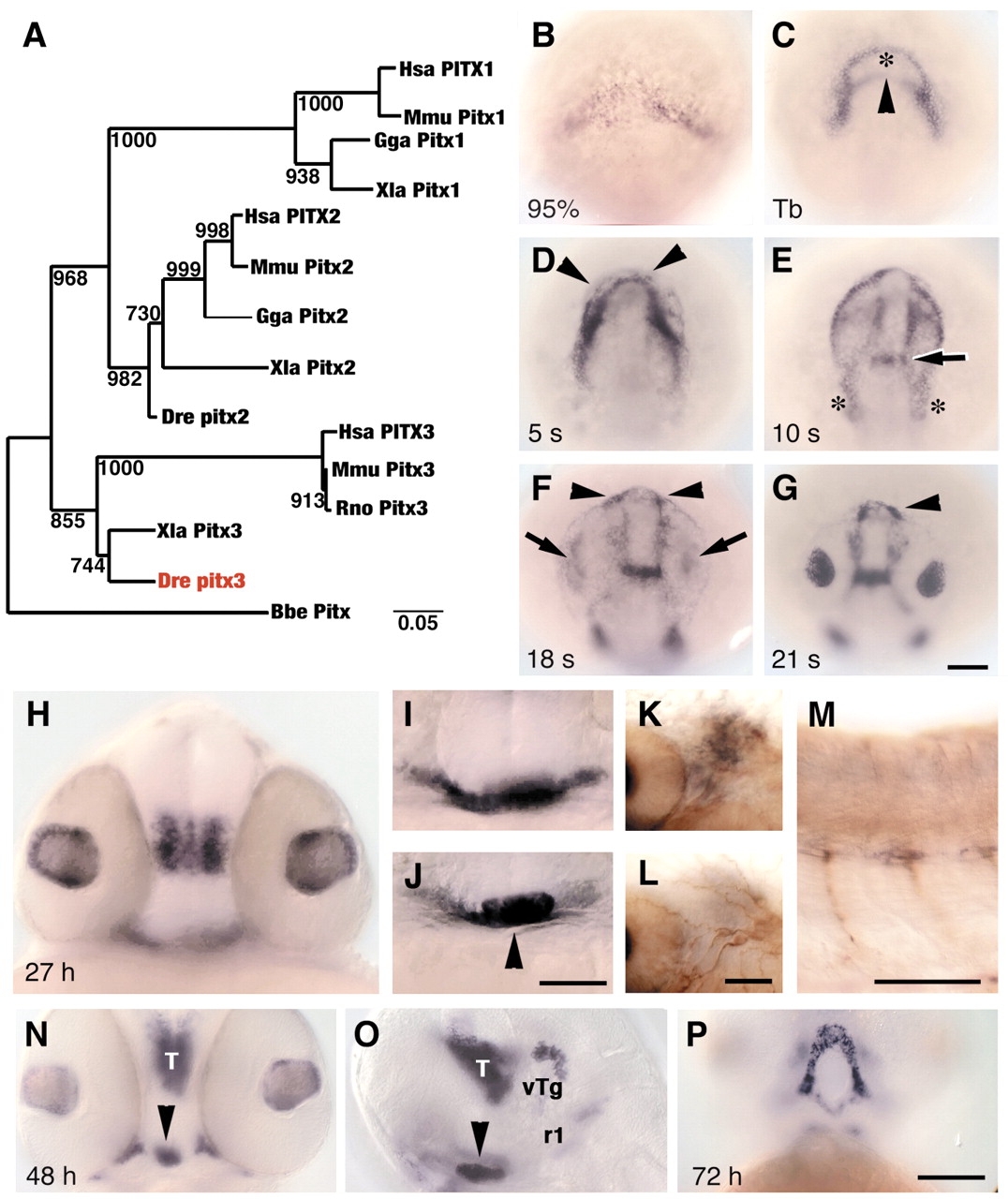
Fig. 1 Zebrafish lens and pituitary cells express Pitx3. (A) Nearest-neighbor joining tree of full-length Pitx amino acid sequences. Zebrafish Pitx3 (red) groups with Xenopus and mammalian Pitx3 proteins. Bootstrap analysis: random number seed, 111; numbers on branches indicate the number of times each node was obtained in 1000 runs. Danio rerio (Dre, AF181681), Homo sapiens (Hsa, P78337, AF238048, AF041339), Mus musculus (Mmu, NM_011097, NM_011098, NM_008852), Rattus norvegicus (Rno, NM_019334, NM_019247), Gallus gallus (Gga, AF069397, AF076640), Xenopus laevis (Xla, AF217647, AF077767, AF297713) and Branchiostoma belcheri (Bbe, AF195616). (B-J) In wild-type embryos, pitx3 is expressed (black) at (B) 95% epiboly; (C) bud stage, in two domains that outline underlying polster (asterisk) and anterior neural plate border (arrowhead); (D) five-somite stage, in cells (arrowheads) around the anterior neural keel; (E) 10-somite stage, presumptive first branchial arch precursors (asterisks) and presumptive forebrain-midbrain border (arrow); (F,G) 18- and 21-somite stages, in prospective lens (arrows) and pituitary placodes (arrowheads); (H) 27 h, in lens and ventral posterior diencephalon; (I) 27 h, in ventral head mesenchyme; (J) 27 h, pituitary (arrowhead). (K-M) 27 h. Double labeling with mRNA probe for pitx3 (black) and anti-acetylated tubulin (brown) indicates that branchial arch mesenchyme (K), which is posterior to eye and ventral to trigeminal placode (L), and trunk muscle pioneer cells (M) express pitx3. (N-P) At 48-72 h, cells in pituitary (arrowheads), thalamus (T), ventral tegmentum (vTg), first rhombomere (r1) and Meckel′s cartilage (P) express pitx3. (B-G) Dorsal view of prospective head region, ventral towards the top; (H-J,N) frontal view, dorsal upwards; (K,L) dorsal view, anterior towards left, different focal planes of same region; (M,O) side view, anterior towards left and dorsal towards top; (P) ventral view, anterior towards top. Scale bars: 100 µm in B-G,H-J,N-P; 50 µm in K-M.
Image
Figure Caption
Figure Data
Acknowledgments
This image is the copyrighted work of the attributed author or publisher, and
ZFIN has permission only to display this image to its users.
Additional permissions should be obtained from the applicable author or publisher of the image.
Full text @ Development

