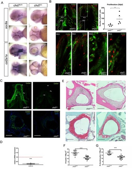
ECM Collagen2a1 deficiency in chd7-/- larvae and adults. (A) WISH of sox9a and col2a1a in craniofacial regions of chd7+/+ and chd7-/- 3dpf larvae, lateral and ventral view (ep: ethmoid plate; pa: palatoquadrate; m: Meckel’s cartilage; ch: ceratohyal; bh: basihyal; cbs: ceratobranchials). (B) Immunohistochemistry for proliferation marker pH 3 (red) and Col2a1 (green) in the craniofacial region of 5dpf chd7+/+(n = 6) and chd7-/- (n = 5) larvae (top; scale bar = 40 μm) and magnified regions of interest (RO1: Region of the Meckel’s cartilage and ceratohyal; RO2: ceratobranchials) (bottom panels; scale bar = 20 μm.) with quantitative analysis of pH 3 positive cells of the craniofacial region (top right panel). (C) Immunofluorescence of Col2a1 in 5 dpf chd7+/+(n = 9) and chd7-/- (n = 8) larvae, ventral view of craniofacial cartilage (top panel; De: dentary; Pq: palatoquadrate; Ch: ceratohyal). Immunofluorescence of Col2a1 in precaudal vertebrae sections of chd7+/+ and chd7-/- mutants with Col2a1 in green and DAPI in blue (bottom panel). (D) RT-qPCR of col2a1a at 9dpf, relative fold change in comparison to chd7+/+ (red dotted line). (E) H&E staining of precaudal vertebrae section of chd7+/+ and chd7-/- mutants (top). Safranin O/fast green staining of Weberian and precaudal vertebrae sections of chd7+/+ and chd7-/- mutants showing cartilage in red (bottom). (F,G) Total nuclei count in Weberian and precaudal vertebral cartilage detected in H&E staining per 100μm2 in chd7+/+ (N = 4, n = 17) and chd7-/- (N = 4, n = 18) and Chondrocyte nuclei count in precaudal vertebral cartilage detected in Safranin O/fast green staining per 100μm2 chd7+/+ (N = 4, n = 18) and chd7-/- (N = 4, n = 19). Scale bar represents 100 μm. Significance: **P < 0.01 ***P < 0.001.
|
