
Fig. 3
- ID
- ZDB-FIG-240517-3
- Publication
- Speirs et al., 2024 - What can we learn about fish neutrophil and macrophage response to immune challenge from studies in zebrafish
- Other Figures
- All Figure Page
- Back to All Figure Page
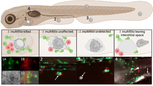
Different scenarios observed in zebrafish larvae infected with the fish parasite Ichthyophthirius multifiliis. 1) Still image from a timelapse video taken with a confocal microscope from Mathiessen et al. (2023) [137]: Neutrophils (1A) and macrophages (1B) are surrounding a parasite, which just died seconds before (1C). 1D is a merge of 1A-C. Images 2–4 have been captured with a stereo microscope: 2) a parasite is surrounded by neutrophils and macrophages but was unaffected for the 5 h it was imaged (white arrow), 3) a parasite is not surrounded by phagocytes (white arrow) and 4) a parasite (white arrow) has left the interstitial space (white arrowhead) which was surrounded by neutrophils and macrophages. Green cells are neutrophils, red cells macrophages and grey cells parasites. |