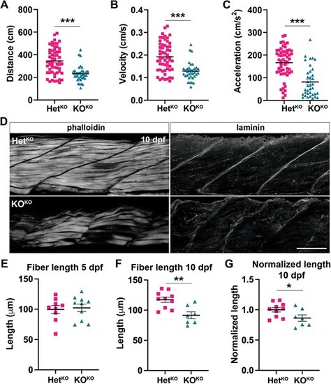
pomt1 KOKO larvae show reduced mobility at 5 dpf and muscle disease at 10 dpf. (A–C) A significant reduction in total distance traveled (A), velocity (B) and acceleration (C) are found in pomt1 KOKO larvae upon automated mobility analysis when compared to HetKO siblings. ***p < 0.001. (D) Staining with fluorescently labeled phalloidin to visualize actin in muscle fibers revealed fiber disorganization from in the 10 dpf KOKO larvae. Laminin staining was used to label the MTJ and showed discontinuities and disruption of the basement membrane between myomeres in KOKOs. See Supplementary Fig. 6A for 5 dpf muscle. Scale bar: 50 μm. (E–G) Fiber length was the same at 5 dpf (E), but it was significantly reduced at 10 dpf (F) even when length was normalized for body length (G). **p < 0.01, *p < 0.05. Alt-text. This figure shows that pomt1 knock-out fish obtained from knock-out mothers have significant locomotor deficits at 5 dpf. In addition, the muscle is severely affected by 10 dpf where we find disorganization of muscle fibers, loss of laminin at the myotendinous junctions, and reduced muscle fiber length.
|
