
Fig. 5
- ID
- ZDB-FIG-240506-65
- Publication
- Lee et al., 2024 - Dysregulated lysosomal exocytosis drives protease-mediated cartilage pathogenesis in multiple lysosomal disorders
- Other Figures
- All Figure Page
- Back to All Figure Page
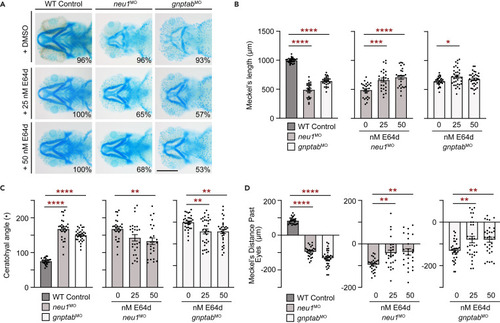
Treatment with the pan cathepsin inhibitor E64d rescues cartilage phenotypes in neu1 and gnptab deficient larvae (A) Alcian blue staining of larvae treated with either 25 nM or 50 nM E64d show inhibiting multiple cathepsin proteases substantially improves cartilage phenotypes in both neu1 and gnptab deficient animals. Percent values represent number of animals exhibiting the pictured phenotype. n = 25–30 larvae per condition from 3 independent experiments. Scale bar: 20 μm. (B) Graphs of Meckel’s length in untreated (graph 1) or E64 treated neu1 (graph 2) or gnptab (graph 3) deficient embryos. (C) Graphs of ceratohyal angle, and (D) Meckel’s distance past the eyes. n = 25–30 embryos per condition from 3 independent experiments. In all cases error = SEM and significance was assessed using the Dunnet’s test (red stars), where ∗p < 0.05, ∗∗p < 0.01, ∗∗∗p < 0.001, and ∗∗∗∗p < 0.0001. |