
Fig. 3
- ID
- ZDB-FIG-240418-7
- Publication
- Hsu et al., 2024 - Macrophages enhance regeneration of lateral line neuromast derived from interneuromast cells through TGF-β in zebrafish
- Other Figures
- All Figure Page
- Back to All Figure Page
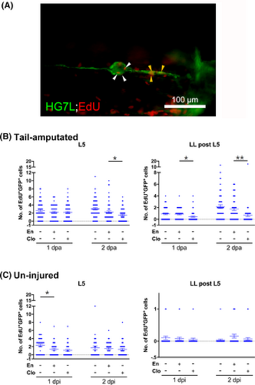
Inhibition of macrophages reduces cell proliferation in the lateral line. We injected encapsome (En) or clodrosomes (Clo) into the posterior cardinal vein of Et(HG7L) larvae 3 days post-fertilization, and the tails were amputated as previously described or left intact. The larvae were fixed 1 and 2 days post-amputation (dpa) and subjected to the EdU assay and immunohistochemistry against EGFP to label proliferating cells (EdU+ in red) and the lateral line (in green), respectively, as shown in a representative image of an untreated larva (a). Then, we counted the number of proliferating lateral line cells around the L5 neuromast (white arrowheads) and the lateral line (LL) behind L5 (yellow arrowheads). (b) For the tail-amputated groups, the treatment effect was significant in both L5 and the LL behind L5 (ANOVA, p < .001). The groups with significant differences as determined by post hoc t-tests are marked on the plots. (c) For the uninjured groups, the treatment effect was significant in the L5 group (ANOVA, p < .01), and only the expression of EdU+ GFP+ cells in the En+/Clo− group at 1 dpi was found to be significantly lower than in the En−/Clo− group (t-test, p < .05). No significant effects were found in the LL behind L5 group (i.e., in between L5 and L6). Error bars indicate SEM. *p < .05; **p < .01. |