
Fig. 3
- ID
- ZDB-FIG-240221-3
- Publication
- Liu et al., 2024 - Live tracking of basal stem cells of the epidermis during growth, homeostasis and injury response in zebrafish
- Other Figures
- All Figure Page
- Back to All Figure Page
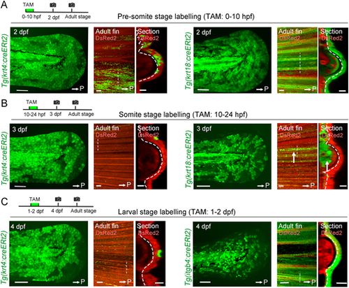
Fate segregation of basal cells from embryonic epithelial cells. (A) Tracking of epithelial cells that are krt4+ or krt18+ in the pre-somite stage. Tamoxifen (TAM) (1 µM) treatment was carried out between 0 and10 hpf. In both Tgs, progenies of EGFP-labelled cells were maintained in the adult fin epidermis including the basal cells and keratinocytes. (B) Tracking of epithelial cells that are krt4+ or krt18+ in the somite stage. EGFP+ cells were induced at 3 dpf (left). In contrast to A, progenies of labelled cells disappeared by the adult stage in both Tgs, except a few remaining surface keratinocytes. In krt18:cre-labelling, a group of mesenchymal cells that were maintained until the adult stage were observed (arrows). (C) Tracking of epithelial cells that are krt4+ or itgb4+ during 1-2 dpf. The progenies of itgb4+ cells contribute to the adult epidermis including the fins (left panels), whereas the progeny of krt4+ cells mostly disappeared by the adult stage (right panels). White arrows indicate the posterior side (P). Respective experimental procedures are schematically shown. Scale bars: 100 µm (2-4 dpf larva and adult fin); 50 µm (section). Straight dashed lines indicate the place of the optical section (right panels). Curved dashed lines indicate basement membrane. |