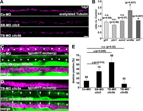
Analyses of Clic5 in ciliogenesis, and in ciliary and glomerular function in zebrafish. (A) Anti-acetylated Tubulin immunostaining reveals reduced cilia formation in the pronephric tubules of embryos injected with SB-MO clic5 (4 ng) or TB-MO clic5b (4 ng) in comparison to Co-MO (4 ng) injected embryos at 1dpf. Representative confocal images depict the middle part of the pronephric tubule for each condition with anterior to the left. Scale bar: 10 µm. (B) Quantitative RT-PCR analyses reveals unaltered expression of Hedgehog signalling components gli1 and ptc1 while Wnt signalling components axin2, wnt8a and lef1 were upregulated upon SB-MO clic5 (4 ng) mediated knockdown compared to the control at 1dpf. (C,D) Injection of TB-MO clic5a (6 ng) leads to detectable FITC-dextran 500 kDa in the pronephric tubules at 4dpf of zebrafish development in comparison to Co-MO (6 ng) injected embryos shown by respective confocal images of Tg(cdh17:mcherry) embryos. Scale bar: 10 µm. (E) Quantification reveals statistic significant FITC-dextran 500 kDa positive embryos for the knockdown of Clic5 and Clic5a compared to the knockdown of Clic5b or the control.
|

