FIGURE

Fig. 3

ID
ZDB-FIG-230825-13
Publication
Dai et al., 2023 - Zebrafish gonad mutant models reveal neuroendocrine mechanisms of brain sexual dimorphism and male mating behaviors of different brain regions
Other Figures
All Figure Page
Back to All Figure Page
Fig. 3

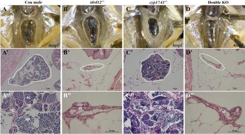
Morphology of primary sex characteristics (PSCs) of the mutants. AA″) Normal testis of a control male. Anatomical view (A); histological view of the whole testis (A′, testis outlined in white); enlarged view shows a well-developed testis with various developmental stages of germ cells, such as spermatocytes (SC), spermatogonia (SG), sperm (SP) and somatic cells (ST) in the testis (A″). BB″ Atrophic testis of a tdrd12−/− fish; the testis lacks germ cells and is surrounded by fatty layers. Anatomical view (B), histological view (B′, outlined in white) and enlarged view (B″). CC″ Testis of a cyp17a1−/− fish. Anatomical view; C histological view (C′, testis outlined in white); enlarged view shows the testis with various developmental stages of germ cells, such as SC, SG, sperm and some ST (C″). DD″ Atrophic testis of a double knockout (KO) fish. Anatomical view (D); histological view (D′, testis outlined in white); enlarged view (D″) shows an atrophic testis similar to that of the tdrd12−/− fish. 6 fish of each genotype were used in this assay

Expression Data

Expression Detail
Antibody Labeling
Phenotype Data

Phenotype Detail
Acknowledgments
This image is the copyrighted work of the attributed author or publisher, and ZFIN has permission only to display this image to its users. Additional permissions should be obtained from the applicable author or publisher of the image. Full text @ Biol Sex Differ