FIGURE

Fig. 3

ID
ZDB-FIG-230613-29
Publication
Čapek et al., 2023 - EmbryoNet: using deep learning to link embryonic phenotypes to signaling pathways
Other Figures
All Figure Page
Back to All Figure Page
Fig. 3

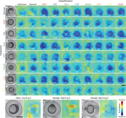
Embryo features activating the neural network.

Class activation heatmaps based on the last convolutional layer of EmbryoNet-Prime showing the part of the image that activates the network at the given timepoint for normal (a) and signaling-defective embryos (bh). Every signaling-defective embryo (−BMP (b), +RA (c), −Wnt (d), −FGF (e), −Nodal (f), −Shh (g), −PCP (h)) is displayed in all classification channels, but only the classification channels corresponding to the correct signaling manipulation show warm colors. The percentages represent the probability of detection. See Supplementary Note 3 for sample sizes. Also see Supplementary Videos 924. ik, Selected embryos showing defects highlighted by the corresponding class activation heatmaps for −Wnt at 13.4 h.p.f. (i) and −Nodal at 10.6 h.p.f. (j) and 26 h.p.f. (k). Scale bars, 500 µm.

Expression Data

Expression Detail
Antibody Labeling
Phenotype Data

Phenotype Detail
Acknowledgments
This image is the copyrighted work of the attributed author or publisher, and ZFIN has permission only to display this image to its users. Additional permissions should be obtained from the applicable author or publisher of the image. Full text @ Nat. Methods