Fig. 2
- ID
- ZDB-FIG-230519-36
- Publication
- He et al., 2022 - Translational control by maternal Nanog promotes oogenesis and early embryonic development
- Other Figures
- All Figure Page
- Back to All Figure Page
|
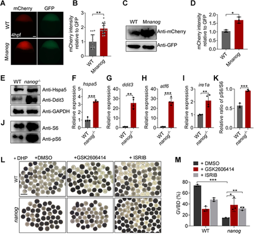
Loss of nanog triggers ER stress and UPR and elevates global translation activity. (A) Fluorescent images showing mCherry reporter levels with GFP protein control levels in WT and Mnanog embryos at 4 hpf. Scale bar: 100 μm. (B) Measurement of mCherry reporter intensities relative to GFP. **P<0.01. n=14. (C,D) Western blotting analysis of mCherry reporter levels at 4 hpf. *P<0.05. (E) Western blot analysis of Hspa5 and Ddit3 in WT and nanog−/− ovaries. (F-I) RT-qPCR analysis of hsp5a (F), ddit3 (G), atf6 (H) and ire1a (I) in WT and nanog−/− ovaries. **P<0.01, ***P<0.001. n=4. (J) Western blot analysis of S6 and phosphorylated S6 in WT and nanog−/− ovaries. The internal control of GAPDH is shown in E. (K) Statistical analysis of phosphorylated S6/S6 ratio shown in J. ***P<0.001. (L) Morphology of stage IV follicles dissected from WT and nanog−/− ovaries and treated with two different PERK inhibitors for 2 h. Follicles dissected from three fish were treated with inhibitors for 2 h (all in the presence of DHP). Final concentrations: GSK2606414, 50 nM; ISRIB, 5 μM; DHP, 1 μg/ml. Scale bar: 1 mm. (M) Comparison of GVBD percentage in WT and nanog mutant follicles treated with or without PERK inhibitor. n=3. *P<0.05, **P<0.01, ***P<0.001. |

