
Fig. 1
- ID
- ZDB-FIG-230322-26
- Publication
- Zhang et al., 2022 - Identification and genetic analysis of rare variants in myosin family genes in 412 Han Chinese congenital heart disease patients
- Other Figures
- All Figure Page
- Back to All Figure Page
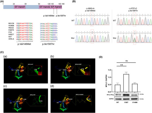
Schematic representation of missense MYL2 variants. (A) MYL2 protein with 166 amino acids showing three EF‐hand domains. The red stars indicate positions of MYL2 variants, and their evolutionary conservation across the species is shown below. (B) DNA sequence chromatogram of wild‐type and mutant variants. The sequences were confirmed by Sanger sequencing. The red squares mark the positions of variants. WT, wild‐type; MUT, mutant. (C) The secondary structure prediction of WT, p. Ile158Thr and p. Val146Met MYL2 was performed using I‐TASSER. (a). MYL2‐WT structure, which emphasized the Ile158 site. The distance between the C‐α of Ser15 and the C‐α of Ile158 is 27.5 Å. (b) MYL2‐I158T structure, which emphasized the Thr158 site. The distance between the C‐α of Ser15 and the C‐α of Thr158 is 30.0 Å, which is longer than the relevant distance in MYL2‐WT. (c). MYL2‐WT structure, which emphasized the Var146 site. The distance between the C‐α of Ser15 and the C‐α of Var146 is 40.4 Å. (d). MYL2‐V146 M structure, which emphasized the Met146 site. The distance between the C‐α of Ser15 and the C‐α of Met146 is 43.1 Å, which is shorter than the relevant distance in MYL2‐WT. (D) Western blot analysis of wild‐type and mutant MYL2 proteins in HEK293T cells. GAPDH was used as a loading control for the cell lysate. ns p > .05, *p < .05, ** < .01. |