
Figure 2
- ID
- ZDB-FIG-230124-151
- Publication
- Grossi et al., 2022 - Lasp1 Expression Is Implicated in Embryonic Development of Zebrafish
- Other Figures
- All Figure Page
- Back to All Figure Page
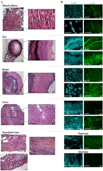
Detection of Lasp1 protein expression in various organs and tissues of zebrafish, including the muscle, the eye, the brain, the liver and the gastrointestinal tract. (A) Histological analysis by hematoxylin and eosin staining of muscle fiber, eye, brain, liver and intestinal tract tissue sections. Pancreas (a), liver (b), and intestine (c) samples are indicated. Magnification: 4× and 20×. (B) Immunofluorescence detection of Lasp1 (green) in the selected tissue sections by immunohistochemistry with Alexa photosensitive antibodies. Magnification: 20× and 63× (blue: DAPI; green: FITC). Scale bars are placed at the lower left corner of each image and correspond to 100 µm for 4× and 20× magnification and to 30 µm for 63× magnification. |
| Gene: | |
|---|---|
| Antibody: | |
| Fish: | |
| Anatomical Terms: | |
| Stage: | Adult |