
Figure 5.
- ID
- ZDB-FIG-230115-44
- Publication
- Bearce et al., 2022 - Urotensin II-related peptides, Urp1 and Urp2, control zebrafish spine morphology
- Other Figures
- All Figure Page
- Back to All Figure Page
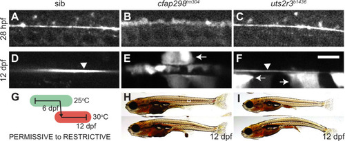
(A–F) Grayscale maximal intensity projection of Sspo-GFP localization in the central canal in 28 hpf embryos (A–C) and 12 dpf adolescents (D–F). RF is denoted by arrow heads in D and F. Arrows point to structures along the central canal that become GFP-positive in cfap298tm304 and uts2r3b1436 mutants. Scale bar: 10 µm. (G) Schematic of temperature shift experiment in which cfap298tm304 mutants are initially raised at permissive temperatures before being shifted to restrictive temperatures at 6 dpf, then imaged at 12 dpf. (H–I) Lateral views of cfap298tm304 (H) and uts2r3b1436 (I) mutants at 12 dpf when Sspo-GFP imaging took place. The white box in H shows the location imaged in D–F.
|
| Gene: | |
|---|---|
| Fish: | |
| Anatomical Terms: | |
| Stage Range: | Prim-5 to Days 7-13 |
| Fish: | |
|---|---|
| Observed In: | |
| Stage Range: | Prim-5 to Days 7-13 |