
Figure 2
- ID
- ZDB-FIG-221226-267
- Publication
- Baniel et al., 2022 - Cutaneous and Developmental Effects of CARD14 Overexpression in Zebrafish
- Other Figures
- All Figure Page
- Back to All Figure Page
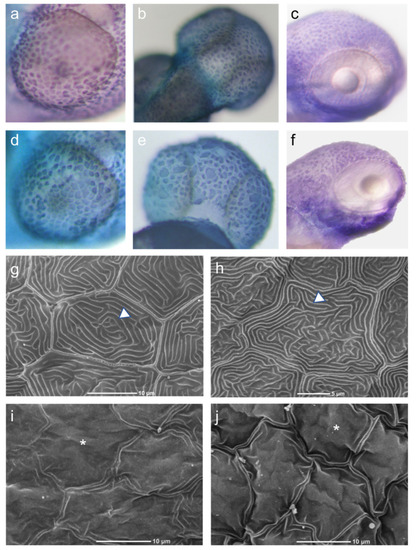
Keratinocyte morphology. Upper panel: Whole mount in situ hybridization with a probe recognizing cytokeratin1 mRNA (dark stain) at 2 dpf (a,c,d,f) and 3 dpf (b,e) demonstrating the distribution and morphology of keratinocytes over the eye (a,d) and over the head (b,c,e–f) areas. Control larvae were injected with an empty plasmid (b) or gfpRNA (a,c), and compared to larvae injected with hCARD14 (e) or zfcard14 (d,f). Note in both regions the uneven distribution and size as well as cell cluster formation as a consequence of CARD14 overexpression as opposed to the even size and regular borders of control keratinocytes. Lower panel: Scanning electron microscopy of 2 dpf embryos. Uninjected (g) and gfp-RNA injected (h) embryos display a uniform and complete array of microridges (triangle), and smooth cell surface, while zfcard14 (i) and hCARD14 injected (j) embryos display uneven cell surface topography (asterisk). |