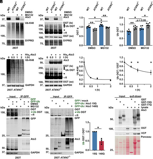
Fig. 3
- ID
- ZDB-FIG-221220-48
- Publication
- Pereira Sena et al., 2021 - Pathophysiological interplay between O-GlcNAc transferase and the Machado-Joseph disease protein ataxin-3
- Other Figures
- All Figure Page
- Back to All Figure Page
|
Ubiquitinated OGT is modulated by ataxin-3. (A and B) Western blot of wild-type (WT) and ataxin-3 knockout (ATXN3−/−) 293T samples. Cells were incubated with DMSO or 10 μM of the proteasome inhibitor MG132 for 4 h prior to harvesting. Proteasome inhibition resulted in increased K48-linked ubiquitin chains (A) and increased full-length (unfilled arrowhead) and high–molecular weight OGT bands (B), interpreted as ubiquitinated OGT (Ub OGT, black arrowheads). 293T ATXN3−/− cells revealed stronger accumulation of Ub OGT. SYPRO Ruby staining served as loading control. fl = full-length; le = long exposure; and se = short exposure. n = 6, one-way ANOVA with Sidak’s post hoc analysis. OGTfl in WT DMSO versus ATXN3−/− DMSO, P = 0.001; in WT DMSO versus MG132, P = 0.029; in ATXN3−/− DMSO versus MG132, P = 0.022; in WT MG132 versus ATXN3−/− MG132, P = 0.004; Ub OGT in WT DMSO versus ATXN3−/− DMSO, P = 0.003; in WT DMSO versus MG132, P = 0.013; and in WT MG132 versus ATXN3−/− MG132, P = 0.016. (C) Western blot of deubiquitination assay. Incubation of 293T ATXN3−/− cell lysates with purified His6-Atx3 resulted in decreased K48-linked ubiquitin chains (Ub-K48) and decreased levels of Ub OGT (black arrowheads) over time. Full-length OGT (unfilled arrowhead) remained unchanged. GAPDH served as loading control. n = 3 to 4, one-sample t test, and Ub OGT in 30, 60, and 120 min incubation with His6-Atx3, P = 0.018, P = 0.018, and P = 0.021 respectively. (D) Western blot of immunoprecipitated GFP and GFP-Ub, probed for OGT. GFP constructs were expressed in WT 293T cells for further lysis and immunoprecipitation (IP) of GFP. Cells were incubated with DMSO or 10 μM MG132 for 4 h prior to lysis. IP of GFP-Ub showed increased full-length and high–molecular weight OGT (Ub OGT, green arrowhead) in cells treated with MG132. Ataxin-3 was used as positive control for GFP-Ub–positive proteins. GAPDH served as loading control. le = long exposure and se = short exposure. (E) Western blot of immunoprecipitated GFP and GFP-Ub from cells expressing Atx3 15Q or 148Q for analyzing Ub OGT. 293T ATXN3−/− cells were transfected with GFP/empty vector (mock) or GFP-Ub/Atx3 (15Q or 148Q) and treated with 10 μM MG132 for 4 h prior to lysis. IP of GFP probed for OGT confirmed the presence of full-length and Ub OGT (green arrowhead) among GFP-Ub–positive proteins. Ub OGT band was weaker in the IP from cells expressing Atx3 148Q. Ataxin-3 was used as positive control for GFP-Ub–positive proteins. GAPDH served as loading control. n = 3, one-sample t test, and P = 0.029. (F) GST pull-down assay for analyzing interaction between ataxin-3 and OGT. GST-tagged WT (GST-15Q) and polyQ-expanded (GST-77Q) ataxin-3 were isolated and incubated with 293T cell lysates. Western blot analysis revealed interaction of both ataxin-3 variants with OGT. GST empty vector was used as negative control for protein interaction and the valosin-containing protein (VCP) was employed as positive control. Total protein was stained with Ponceau S. Data are represented as means ± SEM, *P ≤ 0.05, and **P ≤ 0.01. |

