
Fig. 1
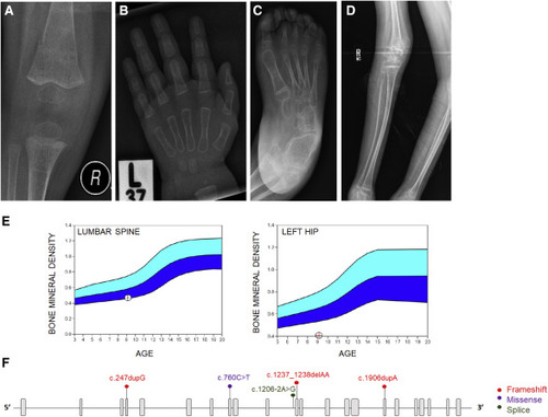
Figure 1. COPB2 variants lead to osteopenia and fractures (A) Right knee radiograph of subject 3 at age 14 months showing fracture of distal femoral metadiaphysis and osteopenia. (B) Left hand radiograph of subject 3 at age 20 months showing thin metacarpal cortices. (C) Right foot radiograph of subject 5 at age 8 years showing diffuse osteopenia and hind-foot varus deformity. (D) Right leg radiographs of subject 6 at age 12 years showing gracile, over-tubulated long bones. (E) Bone densitometry scan in subject 2 showing low bone mineral density of the lumbar spine (left panel) and left hip (right panel). (F) Schematic illustration of COPB2 variants identified in subjects with osteopenia and developmental delay (GenBank: NM_004766.2). |