
Fig. 1
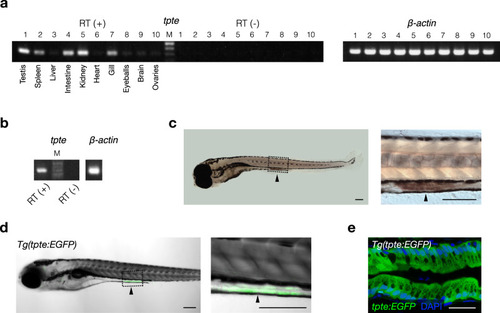
Expression profile of the Dr-Vsp-encoding gene (tpte) in zebrafish tissues.
a RT (+), tpte expression in adult zebrafish tissues: (1) testis, (2) spleen, (3) liver, (4) intestine, (5) kidney, (6) heart, (7) gill, (8) eyeballs, (9) brain, and (10) ovaries. RT (−), negative control for all tissues in the same order. β-actin, positive control. M, marker. b RT (+), tpte expression in 7-dpf zebrafish larva. RT (−), negative control. β-actin, positive control. M, marker. c Whole-mount in situ hybridization (WISH) of 6-dpf zebrafish larva showing tpte-positive signal (arrowhead) in enterocytes of mid-intestine. Scale bar = 200 µm. d A 5-dpf Tg(tpte:EGFP) zebrafish larva expressing EGFP under the promotor of the Dr-Vsp-encoding gene in enterocytes of mid-intestine (arrowhead). The EGFP-positive cells in this figure correspond to the tpte-positive cells in c. Green, Dr-Vsp (tpte:EGFP). Scale bar = 200 µm. e Confocal image of larval enterocytes at mid-intestine of a 5-dpf Tg(tpte:EGFP) zebrafish larva (sagittal section). The enterocytes in this region are known as lysosome-rich enterocytes (LREs) that contain large supranuclear vacuoles. Green, Dr-Vsp (tpte:EGFP). Blue, DAPI. Scale bar = 25 µm. |
| Genes: | |
|---|---|
| Fish: | |
| Anatomical Terms: | |
| Stage Range: | Day 5 to Adult |