
Fig. 18
- ID
- ZDB-FIG-220809-59
- Publication
- Yáñez et al., 2021 - The organization of the zebrafish pallium from a hodological perspective
- Other Figures
- All Figure Page
- Back to All Figure Page
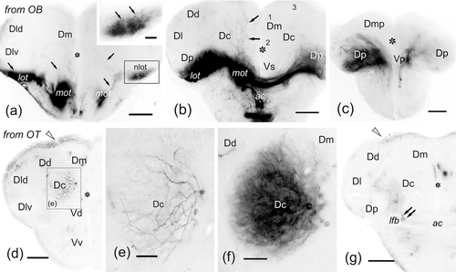
(a–g) Photomicrographs of transverse sections at precommissural (a, d–e) commissural (b, g) and postcommissural (c) levels through telencephalic lobes showing labeled cells (arrowheads) and fibers (arrows) after DiI application to the olfactory bulb (a–c) and rostral optic tectum (d–g). Note the heavy bilateral olfactory projections on the nlot (detailed in a) and Dp in a–c, as well as fibers in Dm at commissural level (b). Note also projections on the lateral region of Vs and Vv (for details of projections, see Gayoso et al., 2011, 2012). (d–g) Note the intense labeling of Dc neurons extending dendritic trees to the central neuropil (d–f) at precommissural levels and sending axons to the optic tectum via the lateral forebrain bundle (g). No labeled cells were observed at commissural telencephalic levels (g). Ipsilateral is to the left. Numbers 1–3 indicate Dm subdivisions. Asterisk, ventricle. For abbreviations, see the list. All photomicrographs are negative images of fluorescent data. Scale bars: 200 μm (a–b, d, g); 100 μm (b); 50 μm (e, inset in a); 25 μm (f) |