FIGURE

Fig. 5.

ID
ZDB-FIG-220703-5
Publication
Zhu et al., 2022 - Computational refocusing of Jones matrix polarization-sensitive optical coherence tomography and investigation of defocus-induced polarization artifacts
Other Figures
All Figure Page
Back to All Figure Page
Fig. 5.

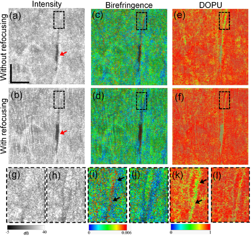
Original (first column) and computationally refocused (second column) en face porcine triceps brachii muscle images. The images are extracted from a depth away from the depth of focus. The first to third columns are the intensity, birefringence, and DOPU images, respectively. (g)-(l) are corresponding magnified images of the regions outlined by black-dashed boxes, where the left images are without refocusing and the right ones are with computational refocusing. Red arrows in (a) and (b) denote a hollow region, which exhibits evident sharpening following computational refocusing. Black arrowheads in (i) and (k) denote birefringence and DOPU artifacts. Scale bar denotes 500 μm.

Expression Data

Expression Detail
Antibody Labeling
Phenotype Data

Phenotype Detail
Acknowledgments
This image is the copyrighted work of the attributed author or publisher, and ZFIN has permission only to display this image to its users. Additional permissions should be obtained from the applicable author or publisher of the image. Full text @ Biomed. Opt. Express