|
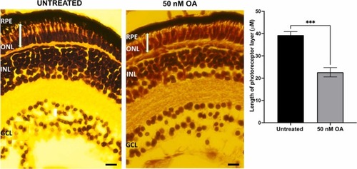
Fig. 7. Haemotoxylin and Eosin (H&E) stained retinal sections of zebrafish embryos untreated and treated with 50 nM OA from 24 to 120hpf. The thickness of the photoreceptor layer (between the RPE layer and the outer plexiform layer, labelled using a white bar) of control and exposed embryos were measured using Image J software. For each condition, the data were collected by measuring five images from sections from four eyes. Data was presented as mean ± SEM (n = 5). * ** p < 0.001. GCL, ganglion cell layer; INL, inner nuclear layer; ONL, outer nuclear layer; RPE, retinal pigment epithelium. Scale bar, 40 µm.
|