
Figure 1
- ID
- ZDB-FIG-220131-59
- Publication
- Kouprianov et al., 2022 - brca2-mutant zebrafish exhibit context- and tissue-dependent alterations in cell phenotypes and response to injury
- Other Figures
- All Figure Page
- Back to All Figure Page
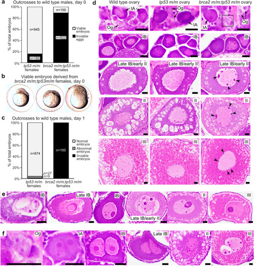
Embryos from brca2 m/m;tp53 m/m female zebrafish undergo proliferation arrest and death and oocytes exhibit abnormal nuclear morphology. (a) Most eggs derived from brca2 m/m;tp53 m/m females are inviable and only a small number of viable embryos are generated, in contrast to tp53 m/m females. Females were outcrossed to fertile wild type males. (b) On day 0, viable embryos derived from brca2 m/m;tp53 m/m embryos are often morphologically abnormal and all undergo developmental arrest at or before sphere stage (approximately four hours post-fertilization). (c) At one day post-fertilization (day 1), greater than 90% of embryos derived from tp53 m/m females are alive and morphologically normal, while all embryos derived from brca2 m/m;tp53 m/m females are dead. (d) Oocytes from brca2 m/m;tp53 m/m females exhibit nuclear abnormalities predominated by aggregated nucleolar material around nuclear margins (black arrowheads). Black box indicates oogonium shown at higher magnification in panel f. (e) Oocytes with massive nucleolar condensation are often degenerate. (f) Infrequent binucleation occurred in oogonia and oocytes from brca2 m/m;tp53 m/m females. Og, oogonia; IA, stage IA oocyte; IB, stage IB oocyte; II, stage II oocyte; III, stage III oocyte. Scale bar = 20 µm. |
| Fish: | |
|---|---|
| Observed In: | |
| Stage Range: | Sphere to Adult |