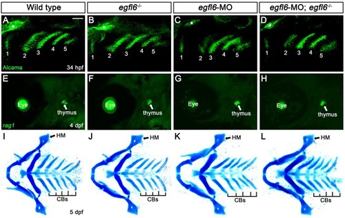
Normal craniofacial development in egfl6 mutants. (A–D) In both wild-type (A, n = 92) and egfl6 mutant (B, n = 31) embryos at 34 hpf, immunohistochemistry for Alcama (green) shows five pouches (1–5). Reduction of egfl6 with a MO in wild-type (C, n = 64) or egfl6 mutant (D, n = 24) animals display normal five pouches. Sensory ganglia are indicated with asterisks. (E-H) Fluorescent in situ hybridization for rag1 (green) at 4 dpf. In both wild-type (C, n = 74) and egfl6 mutant (D, n = 29) zebrafish, rag1 is expressed normally in the thymus. Reduction of egfl6 in wild-type (G, n = 61) or egfl6 mutant (H, n = 17) animals shows normal thymus. (I-L) Ventral whole-mount views of dissected facial cartilages at 5 dpf. Both wild-type (E, n = 105) and egfl6 mutant (F, n = 37) zebrafish invariantly form a triangled shape of hyomandibular (HM) and five ceratobranchial (CB) cartilages on each side. egfl6-MO-injected animals in wild-type (K, n = 88) or egfl6 mutant (H, n = 15) animals have normal facial cartilages, including the HM and CBs. Scale bar: 40 μm.
|

