
FIGURE
Figure 4
- ID
- ZDB-FIG-211002-73
- Publication
- Lu et al., 2021 - Exacerbation of Liver Tumor Metastasis in twist1a+/xmrk+ Double Transgenic Zebrafish following Lipopolysaccharide or Dextran Sulphate Sodium Exposure
- Other Figures
- All Figure Page
- Back to All Figure Page
Figure 4
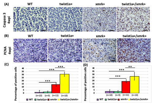
Figure 4. Assessment of apoptosis and cell proliferation in instances of HCC progression in twist1a+/xmrk+ double transgenic zebrafish. Immunohistochemical staining was performed on liver paraffin sections from wild-type, twist1a+, xmrk+, and twist1a+/xmrk+ zebrafish. (A) Caspase-3 staining for apoptosis; and (B) PCNA staining for proliferation at 4 wpi. Scale bar: 50 μm. Quantification of the percentage of cells testing positive for (C) caspase-3 and (D) PCNA. Differences among the variables were assessed using Student’s t-tests. Statistical significance: ** p < 0.01, *** p < 0.001.
|
Expression Data
| Antibodies: | |
|---|---|
| Fish: | |
| Conditions: | |
| Anatomical Term: | |
| Stage: | Adult |
Expression Detail
Antibody Labeling
Phenotype Data
| Fish: | |
|---|---|
| Conditions: | |
| Observed In: | |
| Stage: | Adult |
Phenotype Detail
Acknowledgments
This image is the copyrighted work of the attributed author or publisher, and
ZFIN has permission only to display this image to its users.
Additional permissions should be obtained from the applicable author or publisher of the image.
Full text @ Pharmaceuticals (Basel)