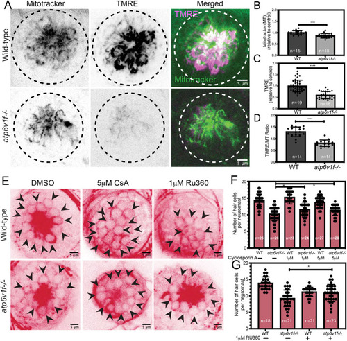
Reduced mitochondrial membrane potential contributes to hair cell death in V-ATPase mutant neuromasts. (A) The vital dyes MitoTracker and TMRE were used to assess mitochondrial mass and mitochondrial transmembrane potential, respectively, in live wild-type and atp6v1f−/− embryos at 4 dpf. Dashed line circles indicate the neuromast boundary. (B,C) Quantification of mean fluorescence intensity measurements per neuromast of MitoTracker (B) and TMRE (C) in wild-type and atp6v1f−/− embryos at 4 dpf. (D) Ratio of TMRE fluorescence intensity to MitoTracker. n=number of embryos. ****P<0.0001 by unpaired Student's t-test with Welch's correction. (E) Representative images of acetylated tubulin immunostaining of hair cells in wild-type and atp6v1f−/− embryos at 4 dpf after treatment with DMSO (vehicle control), CsA or RU360 from 2 dpf to 4 dpf. Arrowheads point to individual hair cells. (F,G) The number of hair cells per neuromast in control embryos and embryos treated with CsA (F) or RU360 (G). n=number of embryos. *P<0.04 by two-way ANOVA with Bonnferroni–Šidák post hoc test.
|
