
Figure 4.3
- ID
- ZDB-FIG-210607-20
- Publication
- Okuda et al., 2021 - Live-imaging of endothelial Erk activity reveals dynamic and sequential signalling events during regenerative angiogenesis
- Other Figures
- All Figure Page
- Back to All Figure Page
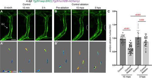
Vessel wounding is required for sustained Erk activity in ablated ISV ECs. (A,B) Vessels that are not wounded do not sustain Erk activity. Lateral spinning disc confocal images of ISV endothelial cells (ECs) in 4 days post-fertilisation (dpf) EC-EKC larvae at 0 min/pre-ablation (left), 15 min/15 min post-ablation (mpa) (middle), or 3 hr/3 hours post-ablation (hpa) (right). Image (A) shows an ISV in a non-ablated control larva and image (B) shows an ISV in a larva with tissue ablated in between two ISVs (control ablation). Images (A) and (B) show fli1aep:EKC expression, while images (A’) and (B’) show the nuclear fli1aep:EKC intensity. White dotted lines show the wounded site. (C) Quantification of post-/pre-ablation nuclear EKC intensity of ECs in either non-ablated control ISVs or control ablation ISVs at 15 mpa (control, 39 ECs, n = 13 larvae; control ablation, 48 ECs, n = 16 larvae) or 3 hpa (control, 18 ECs, n = 6 larvae; control ablation, 24 ECs, n = 8 larvae). ISV: intersegmental vessel. Statistical test: Kruskal-Wallis test was conducted for graph (C). Error bars represent standard deviation. Scale bar: 15 μm. |