
Figure 3
- ID
- ZDB-FIG-210606-162
- Publication
- Rohrer et al., 2021 - Conditional Loss of the Exocyst Component Exoc5 in Retinal Pigment Epithelium (RPE) Results in RPE Dysfunction, Photoreceptor Cell Degeneration, and Decreased Visual Function
- Other Figures
- All Figure Page
- Back to All Figure Page
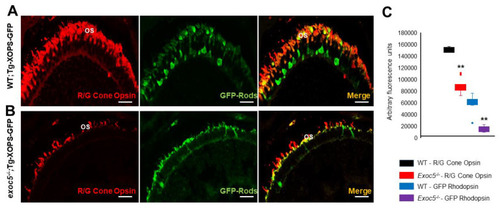
Immunohistochemical analysis of rod and cone photoreceptors in transgenic (Tg:XOPS) wild-type and exoc5 mutant zebrafish. Top panel: wild-type:Transgenic-XOPS (WT;Tg-XOPS-GFP) zebrafish retinas immunostained with R/G cone opsin (red). Bottom panel: exoc5−/− mutant:Transgenic-XOPS (exoc5−/−;Tg-XOPS-GFP) retinas immunostained with R/G cone opsin (red). The transgenic line expresses soluble endogenous GFP in rods (green). Significantly shorter rod and cone OS were observed in exoc5−/− mutant zebrafish, compared to WT zebrafish, at similar ages. (C) Image J was used to quantify fluorescence for R/G cone opsin and GFP+ rods. ** p < 0.005 (WT vs. exoc5−/− mutants). |
| Gene: | |
|---|---|
| Fish: | |
| Anatomical Term: | |
| Stage: | Protruding-mouth |
| Fish: | |
|---|---|
| Observed In: | |
| Stage: | Protruding-mouth |