
Fig. 5
- ID
- ZDB-FIG-210426-45
- Publication
- Wurster et al., 2021 - EGF-mediated suppression of cell extrusion during mucosal damage attenuates opportunistic fungal invasion
- Other Figures
- All Figure Page
- Back to All Figure Page
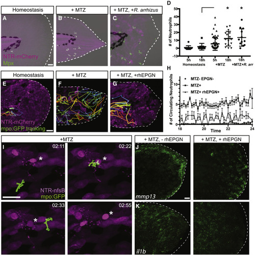
Neutrophils are recruited to sites of mucosal damage and invasive fungal infection (A–C) Maximum intensity projections of confocal images of larvae stained for the neutrophil marker mpx (green) during homeostasis (A), after mucosal damage (B), and mucosal damage and Rhizopus infection. Scale bar, 20 μm. (D) Quantification of the total number of neutrophils present in the epithelium (n = 108 number of animals). (E–G) Still images from time-lapse videos of neutrophil dynamics after cell loss-induced mucosal damage and after treatment with hrEPGN to suppress extrusion. Scale bar, 50 μm. (H) Quantification of the number of circulating neutrophils with and without rhEPGN or rhEGF treatment during induced damage. Data are from 38 animals from three independent experiments. Error bars represent SD. (I) Still images from time-lapse videos of a neutrophil migrating to a site of extrusion (denoted by asterisks). (J and K) Maximum intensity projection images of fluorescent in situ hybridization for mmp13a and il1b in larvae with induced cell loss compared with those treated with hrEPGN. Scale bar, 20 μm. |