
Fig. 1
- ID
- ZDB-FIG-210329-36
- Publication
- Iwasaki et al., 2020 - Development of the anterior lateral line system through local tissue-tissue interactions in the zebrafish head
- Other Figures
- All Figure Page
- Back to All Figure Page
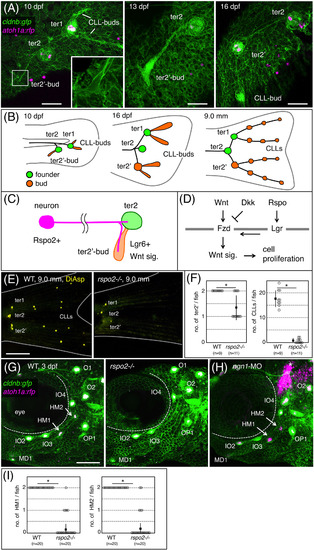
Rspo2 is required for neuromast formation in zebrafish. A, Time‐course observations of the prospective caudal fin region of cldnb:gfp;atoh1a:rfp double transgenic fish at 10, 13, and 16 dpf. Inset shows a higher magnification of the boxed area. B, Schematic drawing for the terminal budding on the caudal fin. C, Proposed model. Rspo2, expressed by sensory neurons, activates Wnt signaling in bud neuromast to promote its proliferation. D, Schematic representation for the regulation of Wnt signaling through its activator (R‐spondin, Rspo) and inhibitor (Dickkopf, Dkk). Fzd, Frizzled; Lgr, Leucine‐rich repeat‐containing G protein‐coupled receptor. E, Caudal fins of wild‐type and rspo2−/− fish stained with DiAsp. F, Quantification of numbers of bud neuromasts in rspo2−/− fish as compared with wild‐type siblings. G, Cranial regions of 3 dpf‐wild‐type and rspo2−/− embryos with cldnb:gfp;atoh1a:rfp background. HM1 and HM2 (arrows in wild‐type) are absent in rspo2−/− embryos. H, 3 dpf‐embryo injected with antisense morpholino against ngn1 (ngn1‐MO). Asterisks indicate the atoh1a:rfp‐expressing hindbrain neurons. I, Quantification of numbers of HM1 and HM2 in rspo2−/− embryos at 4 dpf. Lateral views, anterior is to the left. Data are given as the mean ± SD (F, I) *P < .01 (t‐test), Scale bars: 50 μm (A); 500 μm (E); 100 μm (G) |