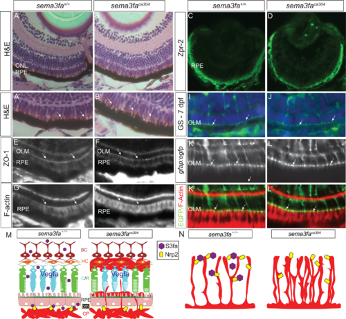
No obvious disruption of the RPE and outer limiting membrane with the loss of Sema3fa. (A, B) Transverse sections of 8 dpf WT and sema3faca304 mutant light-adapted retinas labeled with hematoxylin and eosin. A higher power view of the RPE is shown in A’ and B’. No obvious gaps are seen in the RPE, and apical microvilli (arrows) are evident in both genotypes (N = 3, n = 9 at 72 hpf for both genotypes; N = 2, n = 10 at 8 dpf for both genotypes). (C, D) Immunolabeling of the RPE with the Zpr2 antibody is comparable in WT (N = 2, n = 9) and mutant (N = 2, n = 9) retinas. (E–H) No obvious disruption of the outer limiting membrane (OLM; arrows), as stained with an antibody for the adhesive junction protein ZO-1 (N = 2, n = 10/10 WT eyes; n = 7/7 sema3faca304 eyes) and phalloidin that labels F-actin (N = 3, n = 15/15 WT eyes; n = 16/16 sema3faca304 eyes). (I, J) Glutamine synthetase immunolabeling of the Müller glia in WT (I; N = 2, n = 11/11) and mutant (J: N = 2, n = 10/10) 7 dpf retinas, revealing labeling of the apical Müller glia endfeet at the OLM (arrows) in both genotypes. (K, L) Labeling at the OLM (arrows) of F-actin (phalloidin) and Müller glia endfeet in a Tg(gfap:egfp) background in WT (n = 7/7) and sema3faca304 (n = 8/8) 15 dpf retinas. (M, N) Working model of roles for Sema3fa in regulating vascularization of the neural retina. Choroid vasculature (M): In WT, the choroid plexus (CP) remains outside of the RPE and Bruch's membrane (bm) through the release of Sema3fa (purple hexagon) by cells of the inner nuclear layer and the RPE. If Sema3fa signaling is perturbed, vessels pass through the RPE and leak into the nuclear layers containing the cone and rod (C/R) photoreceptors, horizontal cells (HC), and bipolar cells (BC). Hyaloid/retinal vasculature (N): Sema3fa (purple hexagon) maintains spacing and reduces the density of the blood vessel networks in WT retinas. In the absence of Sema3fa blood vessels overgrow. We propose that in both models Sema3fa acts via Nrp2b (yellow rectangle) and blood vessel growth is supported but not driven by the pro-angiogenic Vegf.
|
