Fig. 2
- ID
- ZDB-FIG-210316-8
- Publication
- Bertozzi et al., 2020 - Is zebrafish heart regeneration "complete"? Lineage-restricted cardiomyocytes proliferate to pre-injury numbers but some fail to differentiate in fibrotic hearts
- Other Figures
- All Figure Page
- Back to All Figure Page
|
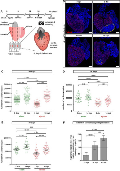
Fig. 2. Zebrafish hearts regenerate all cardiomyocytes lost to injury. (A) Experimental design for quantification of cardiomyocytes. (B) Representative images of −5.1myl7:DsRed2-nlsf2Tg hearts after sham injury or cryoinjury, at 3 or 90 days post surgery. Dashed yellow lines at 3 and 90 dpi outline the wound area. Scale bars, 100 μm. (C) Cardiomyocytes regenerate to the same number as found in sham injured hearts within 90 days. The total number of ventricular cardiomyocytes per heart is plotted. Error bars, CI 95%. n (hearts) = 99 (3 dps), 92 (90 dps), 68 (3 dpi), 58 (90 dpi). One-way ANOVA + Tukey’s multiple comparisons test. Observed difference 90 dpi vs 90 dps = 7400 (4% of CMs in 90 dps); smallest significant detectable difference = 19800 (12%). (D) Cardiomyocyte regeneration is not complete at 14 dpi. n (hearts) = 35 (3 dps), 35 (14 dps), 27 (3 dpi), 31 (14 dpi). One-way ANOVA + Tukey’s multiple comparisons test. (E) Pre-injury cardiomyocyte numbers have been restored at 30 dpi. n (hearts) = 24 (3 dps), 26 (30 dps), 27 (3 dpi), 29 (30 dpi). One-way ANOVA + Tukey’s multiple comparisons test. Observed difference 30 dpi vs 3 dps = 4100 (3.6% of CMs in 3 dps); smallest significant detectable difference = 17600 (15%). (F) The number of regenerated cardiomyocytes at 14, 30 and 90 dpi relative to the number missing in the respective 3 dpi group is plotted. Dotted line represents complete regeneration. n (hearts) = 31 (14 dpi), 29 (30 dpi), 58 (90 dpi). One-way ANOVA + Tukey’s multiple comparisons test. |
Reprinted from Developmental Biology, 471, Bertozzi, A., Wu, C.C., Nguyen, P.D., Vasudevarao, M.D., Mulaw, M.A., Koopman, C.D., de Boer, T.P., Bakkers, J., Weidinger, G., Is zebrafish heart regeneration "complete"? Lineage-restricted cardiomyocytes proliferate to pre-injury numbers but some fail to differentiate in fibrotic hearts, 106-118, Copyright (2020) with permission from Elsevier. Full text @ Dev. Biol.

